
Page 55 - The Netter Collection of Medical Illustrations - Integumentary System_ Volume 4 ( PDFDrive )
P. 55
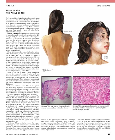
Plate 2-28                                                                                                  Benign Growths

        NEVUS OF OTA
        AND NEVUS OF ITO


        Both nevus of Ota (oculodermal melanocytosis, nevus
        fuscoceruleus  ophthalmomaxillaris)  and  nevus  of  Ito
        (nevus fuscoceruleus acromiodeltoideus) are considered
        to be benign hamartomatous overgrowths of melano-
        cytes. These two processes are located on the face and
        upper  shoulder,  respectively.  They  share  a  common
        pathogenesis and histology with Mongolian spots and
        are  most  likely  caused  by  abnormal  embryological
        migration of melanocytes.                                            Nevus of Ito
          Clinical Findings: The diagnosis of these conditions
        is  most  often  made  on  clinical  grounds,  and  a  skin
        biopsy is rarely, if ever, needed to make the diagnosis.
        Nevus of Ota and nevus of Ito have characteristic loca-
        tions,  and  this  helps  the  clinician  make  the  ultimate
        diagnosis. The closely related Mongolian spot is located
        on  the lower back  of infants  and  manifests as  a  deep
        blue,  asymptomatic  macule  that  almost  always  fades
        away  slowly  until  it  disappears  completely  by  adult-
        hood. It has a higher prevalence in children of Asian or
        Mayan Indian descent.
          Nevus of Ota occurs in a periocular location and can
        affect the bulbar conjunctiva. It is almost always unilat-
        eral in nature.  Nevus of  Ota manifests  as  a  bluish  to                                                          Nevus
        blue-gray macule with indistinct borders that fade into                                                              of Ota
        the  surrounding  normal-colored  skin.  It  is  usually
        located over the distribution of the first two branches
        of  the  trigeminal  nerve.  If  the  bulbar  conjunctiva  is
        involved, the color may vary from bluish gray to dark
        brown. This condition occurs much more commonly in
        women and in patients of Asian descent. Nevus of Ota
        is most often seen in isolation, but on occasion it can
        be seen with a coexisting nevus of Ito.
          Nevus  of  Ito  has  a  similar  clinical  appearance;
        however,  the  location  is  on  the  shoulder  girdle  and
        neck. Unilateral lesions are the rule. The blue to blue-
        gray  macules  can  be  large  and  can  cause  the  patient
        considerable  dismay.  These  lesions  are  asymptomatic
        but can be a major cosmetic concern for patients and
        can  cause  considerable  psychological  and  social
        difficulties.
          Both nevus of Ota and nevus of Ito are more preva-
        lent in the Asian population. Nevus of Ota appears to
        have a very small malignant potential. It is believed that
        Caucasian females with a nevus of Ota are at higher risk
        for transformation into malignant melanoma. Nevus of
        Ito does not appear to have a malignant potential.
          Histology: The histological findings in nevus of Ota,
        nevus  of  Ito,  and  Mongolian  spots  are  identical  and
        resemble those of common blue nevi. Within the lesion,
        nodular  collections  of  melanocytes  are  found  in  the
        dermis, with noticeable elongation of the melanocytes
        in the superficial dermis. There is surrounding fibrosis
        in the dermis with a number of melanophages present.  Nevus of Ota low power. Pigmented mela-  Nevus of Ota high power. Pigmented melanocytes with
          Pathogenesis: Under normal circumstances, melano-  nocytes are spread out within the dermis.  elongated dendritic processes are seen amongst the
        cytes  migrate  during  embryogenesis  from  the  neural                       dermal collagen bundles.
        crest outward to their final locations (e.g., skin, retina).
        Nevus of Ota and nevus of Ito are believed to be caused
        by  abnormal  migration  of  these  melanocytes.  During
        their migration, some unknown signal causes the mela-
        nocytes  to  collect  on  the  face  or  on  the  shoulder,
        respectively.  There  does  not  appear  to  be  a  genetic   Because  of  the  psychological  and  social  hardships   Use of the 1064-nm neodymium:yttrium-aluminum-
        inheritance pattern.                      engendered  by  these  cosmetically  disfiguring  lesions,   garnet (Nd:YAG) laser has resulted in the most success
          Treatment:  These  are  benign  lesions  that  require   therapy  is  appropriate,  albeit  difficult.  If  only  small   in treating these lesions, and it can be used in patients
        no  therapy.  It  is  not  unreasonable  to  monitor  them   areas  are  involved,  cosmetic  makeup  may  be  used  to   of almost any skin type. Q-switching of the laser is a
        clinically for the rare development of malignant trans-  camouflage the region. Topical therapies with hydro-  method  that  has  been  shown  to  increase  its  efficacy.
        formation.  Most  patients  present  for  therapy  because   quinone and tretinoin have shown minimal to no effect   Q-switched  ruby,  alexandrite,  and  1064-nm  Nd:YAG
        they  are  bothered  by  the  appearance  of  the  lesions.   on the pigmentation.  lasers have all been used successfully.


        THE NETTER COLLECTION OF MEDICAL ILLUSTRATIONS                                                                           41