
Page 62 - The Netter Collection of Medical Illustrations - Integumentary System_ Volume 4 ( PDFDrive )
P. 62
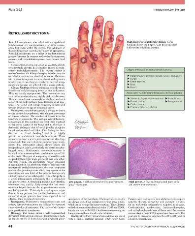
Plate 2-35                                                                                            Integumentary System


       RETICULOHISTIOCYTOMA


       Reticulohistiocytomas,  also  called  solitary  epithelioid                        Multicentric reticulohistiocytomas. Coral
       histiocytomas,  are  conglomerations  of  large  eosino-                           red papules on the fingers. Can be associated
       philic histiocytes within the dermis. The cytoplasm of                             with severe disabling arthritis.
       these cells has been described as “glassy” in appearance.
       Reticulohistiocytomas are a subset of the histiocytoses
       group of diseases. In contrast to the other histiocytoses,
       patients  with  reticulohistiocytoma  have  normal  lipid
       levels.
         Reticulohistiocytomas can occur as a solitary growth
       or as multiple growths in a condition known as multi-
       centric  reticulohistiocytosis.  The  solitary  variant  is                        Organs Involved in Reticulohistiocytoma
       more often seen. On histopathological examination, the
       two clinical variants are identical in nature. Multicen-                             Inflammatory arthritis (hands, knees, shoulders)
       tric reticulohistiocytosis is a rare disease with systemic                           Lungs
       involvement. It can often be a marker of internal malig-                             Bone marrow
       nancy, and patients are afflicted with a severe arthritis.                           Eyes
         Clinical Findings: Solitary lesions are typically small,                           Heart
       firm dermal nodules ranging from 1 to 2 cm in diameter.
       They are usually asymptomatic. Their coloration may                                Associated Autoimmune Diseases and Malignancy
       vary, but most often they are slightly pink to red-brown.
       They are found most commonly on the head and neck                                     Systemic lupus erythematosus  Lymphoma
       region of the body but have been described in all loca-                               Breast cancer             Lung cancer
       tions. They occur with similar frequency in males and                                 Colon cancer
       females and have no age or race predilection.                                         Primary biliary cirrhosis
         Multicentric reticulohistiocytosis is unique in that it
       occurs in an older population, with a higher percentage
       of  females  affected.  The  number  of  lesions  is  in  the
       hundreds to thousands. The multiple reticulohistiocy-
       tomas found in this condition are most often localized
       to  the  dorsal  aspect  of  the  hands  and  to  the  face.  A
       distinctive  finding  is  that  of  small  papules  along  the
       lateral and proximal nail folds. This finding has been
       described  as  “coral  beading,”  and  it  is  highly
       specific  for  multicentric  reticulohistiocytosis.  These
       patients also have a severe arthropathy, and this diag-
       nosis should lead one to look for an underlying malig-
       nancy.  The  arthropathy  almost  always  affects  the
       interphalangeal joints, particularly the distal interpha-
       langeal  joints.  Multicentric  reticulohistiocytosis  is
       believed to be a paraneoplastic condition in up to 25%
       of the cases. The type of malignancy is variable, with
       no  predominant  type  more  prevalent  than  any  other.
       For  this  reason,  age-appropriate  cancer  screening
       is  recommended.  In  about  one  third  of  patients  with
       multicentric  reticulohistiocytosis,  the  joint  symptoms
       precede the growths; in one third, they appear at the
       same time; and one third of the patients develop only
       clinically minor or no arthropathy. This arthropathy is
       a  severe  inflammatory  arthropathy  that  is  symmetric
       and  polyarticular.  A  mutilating  arthritis  may  develop,   Low power. A diffuse dermal infiltrate of “ground-  High power. A few multinucleated giant cells
       sometimes  very  quickly.  Early  recognition  and  treat-  glass” histiocytes      are seen within the tumor.
       ment has helped decrease the progression into severe
       mutilating arthritis. This truly is a multisystem organ
       disease. Many patients have cardiac involvement, and
       almost  all  organ  systems  have  been  reported  to  be
       affected, some with fatal outcomes.       appearance of the cytoplasm. Multinucleate giant cells   Patients with multicentric reticulohistiocytosis require
         Pathogenesis:  Multicentric  reticulohistiocytosis  and   are always seen. They contain more than three nuclei,   systemic  therapy.  Screening  and  constant  vigilance
       solitary reticulohistiocytoma are believed to represent   which can be arranged in many variations. The cells stain   for an underlying malignancy is required in all cases.
       a rare disorder of histiocytes. The cause of the histio-  with the immunohistochemical stains CD45 and CD68,   Corticosteroids,  methotrexate,  hydroxychloroquine,
       cytic proliferation is unknown.           but do not stain with S100. On electron microscopy, no   and cyclophosphamide have all been used. Anti–tumor
         Histology:  The  tumor  shows  a  well-circumscribed   Langerhans cells are found in the infiltrate.  necrosis factor (anti-TNF) agents have been used. The
       dermal infiltrate without a capsule. The infiltrate is made   Treatment: Solitary reticulohistiocytomas are cured   goals are to prevent or suppress the arthropathy and to
       up almost entirely of histiocytes with a “ground-glass”   with  a  simple  elliptical  excision.  They  rarely  recur.   screen for malignancy.

       48                                                                                    THE NETTER COLLECTION OF MEDICAL ILLUSTRATIONS