
Page 57 - The Netter Collection of Medical Illustrations - Integumentary System_ Volume 4 ( PDFDrive )
P. 57
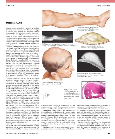
Plate 2-30                                                                                                  Benign Growths


        OSTEOMA CUTIS


                                                                                            Painless bony mass protrudes from
        Osteoma  cutis  is  a  rare  benign  tumor  in  which  bone                         anterior aspect of tibia. Scars due
        formation occurs within the skin. There are two types                               to repeated skin abrasions
        of  osteoma  cutis,  primary  and  secondary.  Primary
        osteoma cutis is idiopathic in nature, whereas secondary
        osteoma cutis is caused by bone formation in an area of
        trauma or another form of cutaneous inflammation. It
        can also be seen secondary to abnormalities of parathy-
        roid  hormone  metabolism,  and  this  form  of  osteoma
        cutis is called metastatic ossification. Secondary osteoma
        cutis  is  much  more  common  than  the  primary  idio-  Radiograph reveals globular outgrowth on tibial
        pathic form.                               cortex with sloping extensions (Codman’s triangles)
          Clinical Findings: Primary osteoma cutis is not asso-                                  Specimen demonstrates continuity
        ciated  with  any  defined  underlying  disorder  and  can                               of tumor with overlying periosteum.
        manifest as a solitary nodule, plaque, or plate-like hard-
        ening of the skin. Some are quite small, whereas others
        are large and cause discomfort. Males and females are
        equally affected, and there is no race predilection. The
        age at onset is variable. Plate-like or plaque-like osteoma
        cutis  is  a  form  of  primary  osteoma  cutis  that  occurs
        during  the  first  few  months  of  life  and  can  even  be
        present at birth. The acral regions are most commonly
        affected.  Over  time,  these  osteomas  tend  to  develop
        ulcerations  or  erosions  of  the  overlying  epidermis.
        With  this  ulceration,  small  parts  of  the  osteoma  are
        extruded  from  the  underlying  dermis  and  expelled
        from the skin. This may be the cause for presentation
        to the clinician. Most patients present with a thickened
        or  hardened  area  of  skin  with  no  preceding  trauma                           Radiograph of excised tumor reveals
        or  inflammatory  condition.  There  is  no  malignant                              densely ossified cortical mass protruding
        potential.                                                                          from outer table of skull.
          Primary  osteomas  of  the  skin  may  be  seen  in  the
        genetically  inherited  disease,  Albright’s  hereditary
        osteodystrophy.  This  condition  is  characterized  by  a   Slowly enlarging, asymptomatic
        constellation  of  findings  including  short  stature,   bony mass on dome of head
        osteoma cutis, mental and physical delay, and brachy-
        dactyly. Varying degrees of obesity and a round appear-
        ance to the face are also seen. This condition is caused
        by an underlying defect in the GNAS gene. This gene                  High power. A well-
        encodes a stimulatory G protein (G s ) that is responsible           circumscribed nodule
        for  cell  signaling  through  the  eventual  production  of         of bone formation just
        cyclic  adenosine  monophosphate  (cAMP).  Albright’s                underneath the epidermis.
        hereditary osteodystrophy has been reported to mani-                 A few haversian canals
        fest with resistance to parathyroid hormone, but other               are present.
        Albright’s  patients  have  not  shown  this  resistance.
        These  differences  are  likely  due  to  the  complex
        inheritance  pattern  and  whether  the  defective  gene
        was  inherited  from  the  maternal  or  paternal  side  or   underlying tissue. This disease is progressive and can   formed by an intramembranous mechanism without the
        both. Most patients have associated hypocalcemia and   result in premature death. This condition is unique in   assistance of a preceding cartilage scaffolding.
        hyperphosphatemia.                        that it is caused by endochondral bone formation.  Treatment: Secondary osteoma cutis can be removed
          Secondary osteoma cutis is far more common than   Pathogenesis: Primary forms of osteoma cutis show   with  a  number  of  surgical  techniques.  Creation  of  a
        the primary form, by a ratio of about 9 : 1. Bone forma-  intramembranous  ossification  that  is  centered  within   small, nick-like incision over the area of osteoma for-
        tion may occur in any area of previous skin trauma, acne   the dermis. There is no preceding cartilage formation   mation and removal with a small curette or laser resur-
        cysts, or epidermal inclusion cysts and are commonly   to act as a scaffolding for the bone to form. The exact   facing has produced the best results. This treatment can
        seen  in  pilomatricomas.  Pilomatricomas  are  benign   cause  is  unknown.  The  G  protein  that  is  defective     be very time-consuming and labor intensive in cases of
        tumors that most often manifest in childhood. Inflam-  in  Albright’s  hereditary  osteodystrophy  has  been     multiple secondary osteoma cutis (e.g., in some cases of
        matory conditions associated with osteoma cutis include   found to be important in bone regulation. The precise   acne-associated osteoma cutis).
        dermatomyositis and scleroderma.          reason why some areas of skin are involved while others   The treatment of primary plaque-like osteoma cutis
          Fibrodysplasia ossificans progressiva is a rare genetic   are  left  intact  in  this  genetic  disease  is  not  well   is surgical removal. Albright’s heriditary osteodystrophy
        condition in which connective tissue is turned into bone   understood.              and  fibrodysplasia  ossificans  progressiva  are  rare  dis-
        after minor trauma, causing secondary osteomas. The   Histology: Areas of bone formation are seen ectopi-  eases  that  require  a  multidisciplinary  approach  at
        skin can be involved, but so can the muscle and other   cally in the dermis or subcutaneous tissue. The bone is   centers with experience treating these conditions.


        THE NETTER COLLECTION OF MEDICAL ILLUSTRATIONS                                                                           43