
Page 67 - The Netter Collection of Medical Illustrations - Integumentary System_ Volume 4 ( PDFDrive )
P. 67
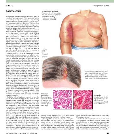
Plate 3-2                                                                                                Malignant Growths


        ANGIOSARCOMA                               Stewart-Treves syndrome.
                                                   Plaque on chronic edematous
                                                   arm. The chronic lymphedema
        Angiosarcoma is a rare, aggressive, malignant tumor of   is secondary to prior
        vascular or lymphatic vessels. These tumors can be seen   mastectomy and axillary
        as  a  solitary  finding  or  secondary  to  long-standing   lymph node dissection.
        lymphedema, such as after radiation therapy or an axil-
        lary or inguinal lymph node dissection. This latter form
        tends to occur years after the radiation or surgical pro-
        cedure. Soft tissue sarcomas are very rare and make up
        a small percentage of all malignancies reported.
          Clinical Findings: Angiosarcomas are most common
        in the older male population. They have no race predi-
        lection. The tumors most commonly arise in the head
        and  neck  region  and  can  manifest  in  many  fashions.
        They often appear as a red to purple plaque with ill-
        defined borders. They can often look like a bruise, and
        the diagnosis can be delayed. The tumor continues to
        expand, forms satellite foci of involvement, and eventu-
        ally ulcerates and bleeds. For some reason, the scalp and
        face of older men are most commonly involved. The
        tumor has a propensity to involve sun-exposed areas of
        the  face  and  scalp.  The  tumors  typically  show  an
        aggressive  growth  pattern  and  have  a  tendency  to
        metastasize early in the course of disease.
          Angiosarcomas can also arise in regions of previous
        long-standing lymphedema caused by radiation expo-
        sure  or  surgical  procedures.  Any  procedure  that  can
        result  in  abnormal  lymphatic  drainage  can  lead  to
        chronic lymphedema. It is believed that long-standing
        lymphedema can result in the development of angiosar-
        coma. Common surgical procedures that cause chronic
        lymphedema are radical mastectomies and lymph node
        dissections  of  the  axilla  or  groin  after  a  diagnosis  of
        lymph  node  involvement  by  breast  cancer  or  mela-
        noma.  Angiosarcomas  arising  in  areas  of  chronic
        lymphedema were first described by Stewart and Treves                                          Angiosarcoma arising from the scalp
        and  have  been  given  the  eponym  Stewart-Treves  syn-                                      of a 65-year-old man. Red indurated
        drome. This type of angiosarcoma is highly aggressive                                          plaque with a central crust due to
        and portends a poor outcome. The Stewart-Treves type                                           underlying ulceration of the tumor.
        of angiosarcoma has been reported most commonly in                                             These tumors can be very aggressive.
        women  who  have  undergone  radical  mastectomy  or
        lymph node dissection for treatment of breast cancer.
        After  years  of  chronic  lymphedema  in  the  ipsilateral
        limb, the patient may develop a reddish, bruise-like area
        on  the  limb.  This  area  slowly  enlarges  and  develops
        plaque-like areas or nodules within the affected region.
        At this point, the diagnosis is often entertained, and the
        diagnosis is made with a skin biopsy. These tumors tend
        to be large at diagnosis, which most likely accounts for   Hemangio-
        the poor prognosis.                       pericytoma.                                                     Hemangio-
          Radiation-induced angiosarcomas may occur at the   Eccentric hyper-                                     endothelioma. Central
        site  of  the  radiation  therapy  or  as  a  result  of  long-  chromatic nuclei                         hyperplastic capillary
        standing chronic lymphedema if the radiation therapy   of pericytic cells                                 surrounded by
        interrupts the lymphatic drainage. These tumors also   surrounding                                        malignant endothelial
        tend to be diagnosed after they have become quite large,   vascular spaces.                               cells. (H&E stain)
        and this portends a poor prognosis. These tumors tend   (H&E stain)
        to occur 4 to 10 years after the initial radiation therapy.
          Pathogenesis: Angiosarcomas are soft tissue tumors
        that  are  derived  from  the  endothelial  lining  of  small
        blood or lymphatic vessels. Some tumors are found to
        have  elevated  levels  of  vascular  endothelial  growth
        factor  (VEGF),  which  is  critical  in  the  regulation  of   radiation  on  the  endothelial  DNA.  No  relation  with   lumina. The same tumor can contain well and poorly
        vessel growth. Other potential players in the pathogen-  human herpesvirus-8 infection has been proven.  differentiated regions.
        esis of this tumor are mast cells, which cause an increase   Histology: All angiosarcomas share the same patho-  Treatment:  The  standard  treatment  is  wide  local
        in stem cell factor; Fas and Fas ligand expression; and   logical features. The tumor lobules are poorly circum-  excision with the goal of obtaining clear margins. This
        lack of the vascular endothelial cadherin protein. All of   scribed and have an infiltrative growth pattern. They   is usually followed by postoperative radiation therapy.
        these factors may interact in an unknown way to induce   contain large amounts of vascular tissue in a disorga-  The  5-year  survival  rate  is  low  (15%-20%).  Tumors
        tumorigenesis.  The  exact  mechanism  of  formation  of   nized  arrangement.  The  lining  of  the  vascular  spaces   that are metastatic or nonoperable can be treated pallia-
        angiosarcoma is unknown. Radiation-induced angiosar-  contains  atypical-appearing  endothelial  cells.  Mitoses   tively  with  various  chemotherapeutic  regimens.  The
        coma may result from a direct mutagenic effect of the   are  frequently  encountered,  as  are  intracytoplasmic   median survival time in these cases is 3 to 6 months.


        THE NETTER COLLECTION OF MEDICAL ILLUSTRATIONS                                                                           53