Page 329 - Clinical Application of Mechanical Ventilation
P. 329

Hemodynamic Monitoring  295


                                                            #
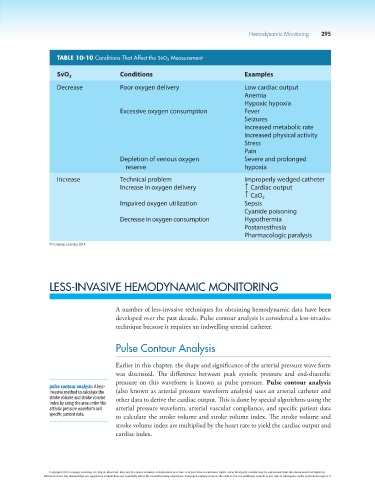
                          TABLE 10-10 Conditions That Affect the SvO 2  Measurement
                            #
                          SvO 2                Conditions                              Examples
                          Decrease             Poor oxygen delivery                    Low cardiac output
                                                                                       Anemia
                                                                                       Hypoxic hypoxia
                                               Excessive oxygen consumption            Fever
                                                                                       Seizures
                                                                                       Increased metabolic rate
                                                                                       Increased physical activity
                                                                                       Stress
                                                                                       Pain
                                               Depletion of venous oxygen              Severe and prolonged
                                                 reserve                               hypoxia

                          Increase             Technical problem                       Improperly wedged catheter
                                               Increase in oxygen delivery               T   Cardiac output
                                                                                          CaO 2
                                                                                         T
                                               Impaired oxygen utilization             Sepsis
                                                                                       Cyanide poisoning
                                               Decrease in oxygen consumption          Hypothermia
                                                                                       Postanesthesia
                                                                                       Pharmacologic paralysis

                        © Cengage Learning 2014


                        LESS-INVASIVE HEMODYNAMIC MONITORING


                                             A number of less-invasive techniques for obtaining hemodynamic data have been
                                             developed over the past decade. Pulse contour analysis is considered a less-invasive
                                             technique because it requires an indwelling arterial catheter.


                                             Pulse Contour Analysis


                                             Earlier in this chapter, the shape and significance of the arterial pressure wave form
                                             was  discussed.  The  difference  between  peak  systolic  pressure  and  end-diastolic
                                             pressure on this waveform is known as pulse pressure. Pulse contour analysis
                        pulse contour analysis: A less-
                        invasive method to calculate the   (also known as arterial pressure waveform analysis) uses an arterial catheter and
                        stroke volume and stroke volume   other data to derive the cardiac output. This is done by special algorithms using the
                        index by using the area under the
                        arterial pressure waveform and   arterial pressure waveform, arterial vascular compliance, and specific patient data
                        specific patient data.  to calculate the stroke volume and stroke volume index. The stroke volume and
                                             stroke volume index are multiplied by the heart rate to yield the cardiac output and
                                             cardiac index.


                        Copyright 2013 Cengage Learning. All Rights Reserved. May not be copied, scanned, or duplicated, in whole or in part. Due to electronic rights, some third party content may be suppressed from the eBook and/or eChapter(s).
                      Editorial review has deemed that any suppressed content does not materially affect the overall learning experience. Cengage Learning reserves the right to remove additional content at any time if subsequent rights restrictions require it.
   324   325   326   327   328   329   330   331   332   333   334