
Page 324 - Clinical Application of Mechanical Ventilation
P. 324
290 Chapter 10
a
c v
y © Cengage Learning 2014
x
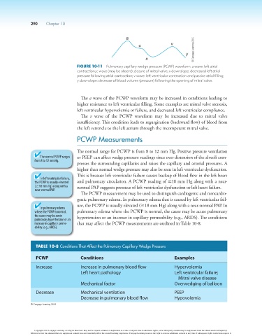
Figure 10-11 Pulmonary capillary wedge pressure (PCWP) waveform. a wave: left atrial
contraction; c wave (may be absent): closure of mitral valve; x downslope: decreased left atrial
pressure following atrial contraction; v wave: left ventricular contraction and passive atrial filling;
y downslope: decrease of blood volume (pressure) following the opening of mitral valve.
The a wave of the PCWP waveform may be increased in conditions leading to
higher resistance to left ventricular filling. Some examples are mitral valve stenosis,
left ventricular hypervolemia or failure, and decreased left ventricular compliance.
The v wave of the PCWP waveform may be increased due to mitral valve
insufficiency. This condition leads to regurgitation (backward flow) of blood from
the left ventricle to the left atrium through the incompetent mitral valve.
PCWP Measurements
The normal range for PCWP is from 8 to 12 mm Hg. Positive pressure ventilation
The normal PCWP ranges or PEEP can affect wedge pressure readings since over distension of the alveoli com-
from 8 to 12 mm Hg.
presses the surrounding capillaries and raises the capillary and arterial pressures. A
higher than normal wedge pressure may also be seen in left ventricular dysfunction.
This is because left ventricular failure causes backup of blood flow in the left heart
In left ventricular failure,
the PCWP is usually elevated and pulmonary circulation. A PCWP reading of ≥18 mm Hg along with a near-
(≥18 mm Hg) along with a normal PAP suggests presence of left ventricular dysfunction or left heart failure.
near-normal PAP.
The PCWP measurement may be used to distinguish cardiogenic and noncardio-
genic pulmonary edema. In pulmonary edema that is caused by left ventricular fail-
ure, the PCWP is usually elevated (≥18 mm Hg) along with a near-normal PAP. In
In pulmonary edema
where the PCWP is normal, pulmonary edema where the PCWP is normal, the cause may be acute pulmonary
the cause may be acute hypertension or an increase in capillary permeability (e.g., ARDS). The conditions
pulmonary hypertension or an
increase in capillary perme- that may affect the PCWP measurements are outlined in Table 10-8.
ability (e.g., ARDS).
TABLE 10-8 Conditions That Affect the Pulmonary Capillary Wedge Pressure
PCWP Conditions Examples
Increase Increase in pulmonary blood flow Hypervolemia
Left heart pathology Left ventricular failure;
Mitral valve disease
Mechanical factor Overwedging of balloon
Decrease Mechanical ventilation PEEP
Decrease in pulmonary blood flow Hypovolemia
© Cengage Learning 2014
Copyright 2013 Cengage Learning. All Rights Reserved. May not be copied, scanned, or duplicated, in whole or in part. Due to electronic rights, some third party content may be suppressed from the eBook and/or eChapter(s).
Editorial review has deemed that any suppressed content does not materially affect the overall learning experience. Cengage Learning reserves the right to remove additional content at any time if subsequent rights restrictions require it.