
Page 332 - Clinical Application of Mechanical Ventilation
P. 332
298 Chapter 10
A B
5 cm
Constant Current
Current Sensing
© Cengage Learning 2014
5 cm
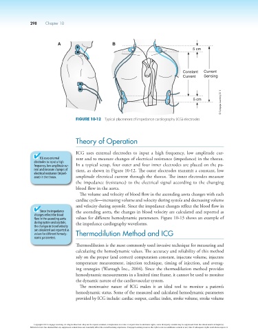
Figure 10-12 Typical placement of impedance cardiography (ICG) electrodes
Theory of Operation
ICG uses external electrodes to input a high frequency, low amplitude cur-
ICG uses external rent and to measure changes of electrical resistance (impedance) in the thorax.
electrodes to input a high
frequency, low amplitude cur- In a typical setup, four outer and four inner electrodes are placed on the pa-
rent and measure changes of tient, as shown in Figure 10-12. The outer electrodes transmit a constant, low
electrical resistance (imped-
ance) in the thorax. amplitude electrical current through the thorax. The inner electrodes measure
the impedance (resistance) to the electrical signal according to the changing
blood flow in the aorta.
The volume and velocity of blood flow in the ascending aorta changes with each
cardiac cycle—increasing volume and velocity during systole and decreasing volume
and velocity during asystole. Since the impedance changes reflect the blood flow in
Since the impedance the ascending aorta, the changes in blood velocity are calculated and reported as
changes reflect the blood
flow in the ascending aorta values for different hemodynamic parameters. Figure 10-13 shows an example of
during systole and asystole, the impedance cardiography waveforms.
the changes in blood velocity
are calculated and reported as
values for different hemody- Thermodilution Method and ICG
namic parameters.
Thermodilution is the most commonly used invasive technique for measuring and
calculating the hemodynamic values. The accuracy and reliability of this method
rely on the proper (and correct) computation constant, injectate volume, injectate
temperature measurement, injection technique, timing of injection, and averag-
ing strategies (Wantagh Inc., 2004). Since the thermodilution method provides
hemodynamic measurements in a limited time frame, it cannot be used to monitor
the dynamic nature of the cardiovascular system.
The noninvasive nature of ICG makes it an ideal tool to monitor a patient’s
hemodynamic status. Some of the measured and calculated hemodynamic parameters
provided by ICG include: cardiac output, cardiac index, stroke volume, stroke volume
Copyright 2013 Cengage Learning. All Rights Reserved. May not be copied, scanned, or duplicated, in whole or in part. Due to electronic rights, some third party content may be suppressed from the eBook and/or eChapter(s).
Editorial review has deemed that any suppressed content does not materially affect the overall learning experience. Cengage Learning reserves the right to remove additional content at any time if subsequent rights restrictions require it.