
Page 334 - Clinical Application of Mechanical Ventilation
P. 334
300 Chapter 10
Accuracy of ICG
Many studies have been done to compare and validate the accuracy of ICG with
The correlation of cardiac other methods of hemodynamic monitoring (Drazner et al., 2002; Ziegler et al.,
output determined by ICG ver-
sus the Fick method and the 1999). In a study of patients with pulmonary arterial hypertension, the correlation of
thermodilution method were cardiac output determined by ICG versus the Fick method and the thermodilution
0.84 and 0.80, respectively.
method were 0.84 and 0.80, respectively (Yung et al., 2004). These correlation
indices are similar to the results of other studies using different patient populations.
Since ICG is less variable and more reproducible than other invasive methods, it has
shown sufficient clinical usefulness to become a standard practice in noninvasive
Technical and measure- hemodynamic evaluations (Van De Water et al., 2003).
ment errors of ICG include:
wrong placement of elec- Methodology Errors. While ICG is useful in many clinical situations, there are some
trodes, abnormal body struc-
ture, tachycardia, presence technical reasons and conditions that may influence the use and accuracy of ICG
of pacemaker, arrhythmias,
open-heart or aorta surgery, (Braždžionytė et al., 2004a). They include wrong placement of electrodes; abnormal
abnormal cardiac anatomy, body structure (cachetic or obese); tachycardia (.120/min); presence of pacemaker;
abnormal hematocrit, and
pleural effusion. arrhythmias; open-heart or aorta surgery; abnormal cardiac anatomy (e.g., transpo-
sitions, aneurysms); abnormal hematocrit; and pleural effusion.
Clinical Application
With ICG, the therapeutic effects of fluid administration and resuscitation can
ICG provides these be assessed by monitoring the stroke volume and cardiac output. ICG has also
advantages: noninvasive
continuous monitoring, rapid been used to evaluate the hemodynamic status of critically ill patients in the inten-
diagnosis and assessment of sive care units, surgical areas, and outpatient and emergency departments (Bishop
cardiopulmonary status,
hemodynamic response to et al., 1996; Milzman et al., 1997; Shoemaker et al., 1994; Wo et al., 1995; Yancey,
fluids and drugs, and availability 2003). Evaluation and follow-up of patients with acute myocardial infarction is also
outside the critical care area.
possible with ICG (Braždžionytė et al., 2004b).
In subacute care, adjustment of the dosages of cardiovascular drugs can be done
by monitoring the thoracic fluid status, stroke volume, and cardiac output (Franz,
1996). Since ICG monitoring is noninvasive, it can be used in outpatients as well as
patients at home. Some advantages of ICG are listed in Table 10-12.
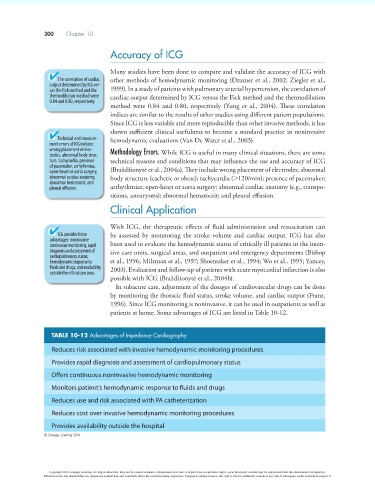
TABLE 10-12 Advantages of Impedance Cardiography
Reduces risk associated with invasive hemodynamic monitoring procedures
Provides rapid diagnosis and assessment of cardiopulmonary status
Offers continuous noninvasive hemodynamic monitoring
Monitors patient’s hemodynamic response to fluids and drugs
Reduces use and risk associated with PA catheterization
Reduces cost over invasive hemodynamic monitoring procedures
Provides availability outside the hospital
© Cengage Learning 2014
Copyright 2013 Cengage Learning. All Rights Reserved. May not be copied, scanned, or duplicated, in whole or in part. Due to electronic rights, some third party content may be suppressed from the eBook and/or eChapter(s).
Editorial review has deemed that any suppressed content does not materially affect the overall learning experience. Cengage Learning reserves the right to remove additional content at any time if subsequent rights restrictions require it.