
Page 420 - Clinical Application of Mechanical Ventilation
P. 420
386 Chapter 12
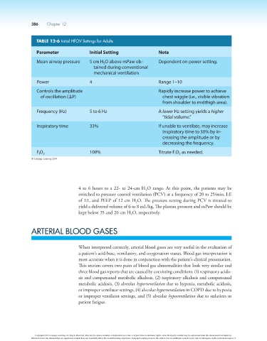
TABLE 12-6 Initial HFOV Settings for Adults
Parameter Initial Setting Note
Mean airway pressure 5 cm H O above mPaw ob- Dependent on power setting.
2
tained during conventional
mechanical ventilation
Power 4 Range 1–10
Controls the amplitude Rapidly increase power to achieve
of oscillation (DP) chest wiggle (i.e., visible vibration
from shoulder to midthigh area).
Frequency (Hz) 5 to 6 Hz A lower Hz setting yields a higher
“tidal volume.”
Inspiratory time 33% If unable to ventilate, may increase
inspiratory time to 50% by in-
creasing the amplitude or by
decreasing the frequency.
F O 2 100% Titrate F O as needed.
I
I
2
© Cengage Learning 2014
4 to 6 hours to a 22- to 24-cm H O range. At this point, the patients may be
2
switched to pressure control ventilation (PCV) at a frequency of 20 to 25/min, I:E
of 1:1, and PEEP of 12 cm H O. The pressure setting during PCV is titrated to
2
yield a delivered volume of 6 to 8 mL/kg. The plateau pressure and mPaw should be
kept below 35 and 20 cm H O, respectively.
2
ARTERIAL BLOOD GASES
When interpreted correctly, arterial blood gases are very useful in the evaluation of
a patient’s acid-base, ventilatory, and oxygenation status. Blood gas interpretation is
most accurate when it is done in conjunction with the patient’s clinical presentation.
This section covers two pairs of blood gas abnormalities that look very similar and
three blood gas reports that are caused by coexisting conditions: (1) respiratory acido-
sis and compensated metabolic alkalosis, (2) respiratory alkalosis and compensated
metabolic acidosis, (3) alveolar hyperventilation due to hypoxia, metabolic acidosis,
or improper ventilator settings, (4) alveolar hyperventilation in COPD due to hypoxia
or improper ventilator settings, and (5) alveolar hypoventilation due to sedatives or
patient fatigue.
Copyright 2013 Cengage Learning. All Rights Reserved. May not be copied, scanned, or duplicated, in whole or in part. Due to electronic rights, some third party content may be suppressed from the eBook and/or eChapter(s).
Editorial review has deemed that any suppressed content does not materially affect the overall learning experience. Cengage Learning reserves the right to remove additional content at any time if subsequent rights restrictions require it.