
Page 498 - Clinical Application of Mechanical Ventilation
P. 498
464 Chapter 14
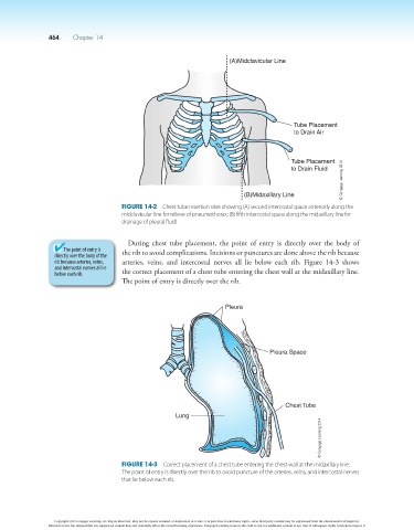
(A)Midclavicular Line
Tube Placement
to Drain Air
Tube Placement
© Cengage Learning 2014
to Drain Fluid
(B)Midaxillary Line
Figure 14-2 Chest tube insertion sites showing (A) second intercostal space anteriorly along the
midclavicular line for relieve of pneumothorax; (B) fifth intercostal space along the midaxillary line for
drainage of pleural fluid.
During chest tube placement, the point of entry is directly over the body of
The point of entry is the rib to avoid complications. Incisions or punctures are done above the rib because
directly over the body of the
rib because arteries, veins, arteries, veins, and intercostal nerves all lie below each rib. Figure 14-3 shows
and intercostal nerves all lie the correct placement of a chest tube entering the chest wall at the midaxillary line.
below each rib.
The point of entry is directly over the rib.
Pleura
Pleura Space
Chest Tube
Lung
© Cengage Learning 2014
Figure 14-3 Correct placement of a chest tube entering the chest wall at the midaxillary line.
The point of entry is directly over the rib to avoid puncture of the arteries, veins, and intercostal nerves
that lie below each rib.
Copyright 2013 Cengage Learning. All Rights Reserved. May not be copied, scanned, or duplicated, in whole or in part. Due to electronic rights, some third party content may be suppressed from the eBook and/or eChapter(s).
Editorial review has deemed that any suppressed content does not materially affect the overall learning experience. Cengage Learning reserves the right to remove additional content at any time if subsequent rights restrictions require it.