
Page 499 - Clinical Application of Mechanical Ventilation
P. 499
Procedures Related to Mechanical Ventilation 465
Methods of Placement
Operative tube thoracostomy and trocar tube thoracostomy are two common
methods to perform chest tube placement. Each method has its advantages and
disadvantages (Deshpande et al., 2002).
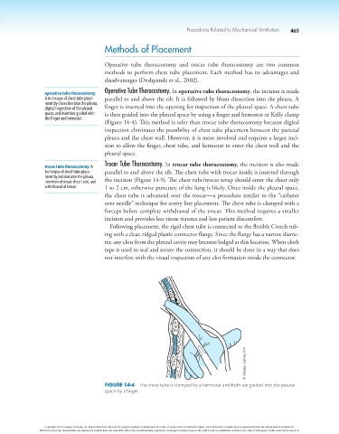
operative tube thoracostomy: Operative Tube Thoracostomy. In operative tube thoracostomy, the incision is made
A technique of chest tube place- parallel to and above the rib. It is followed by blunt dissection into the pleura. A
ment by dissection into the pleura,
digital inspection of the pleural finger is inserted into the opening for inspection of the pleural space. A chest tube
space, and insertion guided with is then guided into the pleural space by using a finger and hemostat or Kelly clamp
the finger and hemostat.
(Figure 14-4). This method is safer than trocar tube thoracostomy because digital
inspection eliminates the possibility of chest tube placement between the parietal
pleura and the chest wall. However, it is more involved and requires a larger inci-
sion to allow the finger, chest tube, and hemostat to enter the chest wall and the
pleural space.
trocar tube thoracostomy: A Trocar Tube Thoracostomy. In trocar tube thoracostomy, the incision is also made
technique of chest tube place- parallel to and above the rib. The chest tube with trocar inside is inserted through
ment by incision into the pleura,
insertion of trocar chest tube, and the incision (Figure 14-5). The chest tube/trocar setup should enter the chest only
withdrawal of trocar. 1 to 2 cm, otherwise puncture of the lung is likely. Once inside the pleural space,
the chest tube is advanced over the trocar—a procedure similar to the “catheter
over needle” technique for artery line placement. The chest tube is clamped with a
forceps before complete withdrawal of the trocar. This method requires a smaller
incision and provides less tissue trauma and less patient discomfort.
Following placement, the rigid chest tube is connected to the flexible Creech tub-
ing with a clear, ridged plastic connector flange. Since the flange has a narrow diame-
ter, any clots from the pleural cavity may become lodged at this location. When cloth
tape is used to seal and secure the connection, it should be done in a way that does
not interfere with the visual inspection of any clot formation inside the connector.
© Cengage Learning 2014
Figure 14-4 The chest tube is clamped by a hemostat and both are guided into the pleural
space by a finger.
Copyright 2013 Cengage Learning. All Rights Reserved. May not be copied, scanned, or duplicated, in whole or in part. Due to electronic rights, some third party content may be suppressed from the eBook and/or eChapter(s).
Editorial review has deemed that any suppressed content does not materially affect the overall learning experience. Cengage Learning reserves the right to remove additional content at any time if subsequent rights restrictions require it.