
Page 500 - Clinical Application of Mechanical Ventilation
P. 500
466 Chapter 14
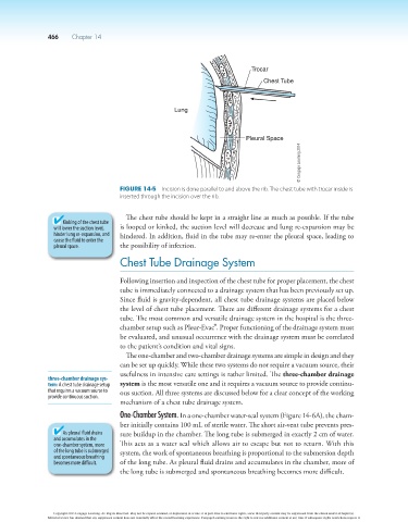
Trocar
Chest Tube
Lung
Pleural Space
© Cengage Learning 2014
Figure 14-5 Incision is done parallel to and above the rib. The chest tube with trocar inside is
inserted through the incision over the rib.
The chest tube should be kept in a straight line as much as possible. If the tube
Kinking of the chest tube
will lower the suction level, is looped or kinked, the suction level will decrease and lung re-expansion may be
hinder lung re-expansion, and hindered. In addition, fluid in the tube may re-enter the pleural space, leading to
cause the fluid to enter the
pleural space. the possibility of infection.
Chest Tube Drainage System
Following insertion and inspection of the chest tube for proper placement, the chest
tube is immediately connected to a drainage system that has been previously set up.
Since fluid is gravity-dependent, all chest tube drainage systems are placed below
the level of chest tube placement. There are different drainage systems for a chest
tube. The most common and versatile drainage system in the hospital is the three-
chamber setup such as Pleur-Evac®. Proper functioning of the drainage system must
be evaluated, and unusual occurrence with the drainage system must be correlated
to the patient’s condition and vital signs.
The one-chamber and two-chamber drainage systems are simple in design and they
can be set up quickly. While these two systems do not require a vacuum source, their
usefulness in intensive care settings is rather limited. The three-chamber drainage
three-chamber drainage sys-
tem: A chest tube drainage setup system is the most versatile one and it requires a vacuum source to provide continu-
that requires a vacuum source to ous suction. All three systems are discussed below for a clear concept of the working
provide continuous suction.
mechanism of a chest tube drainage system.
One-Chamber System. In a one-chamber water-seal system (Figure 14-6A), the cham-
ber initially contains 100 mL of sterile water. The short air-vent tube prevents pres-
As pleural fluid drains sure buildup in the chamber. The long tube is submerged in exactly 2 cm of water.
and accumulates in the
one-chamber system, more This acts as a water seal which allows air to escape but not to return. With this
of the long tube is submerged system, the work of spontaneous breathing is proportional to the submersion depth
and spontaneous breathing
becomes more difficult. of the long tube. As pleural fluid drains and accumulates in the chamber, more of
the long tube is submerged and spontaneous breathing becomes more difficult.
Copyright 2013 Cengage Learning. All Rights Reserved. May not be copied, scanned, or duplicated, in whole or in part. Due to electronic rights, some third party content may be suppressed from the eBook and/or eChapter(s).
Editorial review has deemed that any suppressed content does not materially affect the overall learning experience. Cengage Learning reserves the right to remove additional content at any time if subsequent rights restrictions require it.