
Page 503 - Clinical Application of Mechanical Ventilation
P. 503
Procedures Related to Mechanical Ventilation 469
With a three-chamber drainage system, the fluid collection chamber should be
inspected to note the volume and characteristics of the fluid drainage. The volume
collected should decrease over time.
Care and Removal of Chest Tube
Emergencies may happen to the chest tube setup. If the drainage holes on the chest
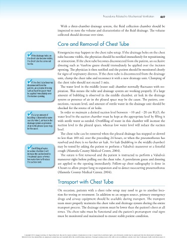
If the drainage holes on tube become visible, the physician should be notified immediately for repositioning
the chest tube become visible,
the chest tube has come out or reinsertion. If the chest tube becomes disconnected from the patient, an occlusive
too far. dressing such as Vaseline gauze should immediately be applied over the incision
opening. The physician is then notified and the patient should be monitored closely
for signs of respiratory distress. If the chest tube is disconnected from the drainage
unit, clamp the chest tube and reconnect it with a new drainage unit. Clamping of
If the chest tube becomes the chest tube should not exceed 1 min.
disconnected from the The water level in the middle (water seal) chamber normally fluctuates with res-
patient, an occlusive dressing
such as Vaseline gauze must piration. This means the tube and drainage system are working properly. If a large
be applied immediately over
the incision opening. amount of bubbling is observed in the middle chamber, air leak in the drainage
system or presence of air in the pleural space may be the cause. The patient, con-
nections, vacuum level, and amount of sterile water in the drainage unit should be
checked for the source of air leaks.
In order to maintain a desired suction level between 210 and 220 cm H O, the
2
If a large amount of
bubbling is observed in water water level in the suction chamber must be kept at the appropriate level by filling it
seal chamber 2, air leak in the with sterile water as needed. Overfilling of water in this chamber will increase the
drainage system or presence
of air in the pleural space may suction level to the pleural space, whereas low water level will reduce the suction
be the cause. level.
The chest tube can be removed when the pleural drainage has stopped or slowed
to less than 100 mL over the preceding 24 hours, or when the pneumothorax has
resolved and there is no further air leak. Air leak (bubbling in the middle chamber)
may be tested by asking the patient to perform a Valsalva’s maneuver or a forceful
Overfilling of water
in suction chamber 3 will cough (Alameda County Medical Center, 2004).
increase the suction level to The suture is first removed and the patient is instructed to perform a Valsalva’s
the pleural space, whereas
low water level will reduce maneuver right before pulling out the chest tube. A petrolatum gauze and dressing
the suction level. are applied to the opening immediately. Follow-up chest radiography is done in
4 hours to allow proper lung re-expansion and to detect reoccurring pneumothorax
(Alameda County Medical Center, 2004).
Transport with Chest Tube
On occasion, patients with a chest tube setup may need to go to another loca-
tion for testing or treatment. In addition to an oxygen source, primary emergency
drugs and airway equipment should be available during transport. The transport
team must properly maintain the chest tube and drainage system during the entire
transport process. The drainage system must be lower than the patient’s chest at all
times. The chest tube must be functional and the patient’s pretransport vital signs
must be monitored and maintained to ensure stable patient condition.
Copyright 2013 Cengage Learning. All Rights Reserved. May not be copied, scanned, or duplicated, in whole or in part. Due to electronic rights, some third party content may be suppressed from the eBook and/or eChapter(s).
Editorial review has deemed that any suppressed content does not materially affect the overall learning experience. Cengage Learning reserves the right to remove additional content at any time if subsequent rights restrictions require it.