
Page 69 - Clinical Application of Mechanical Ventilation
P. 69
Effects of Positive Pressure Ventilation 35
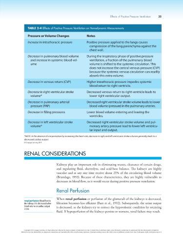
TABLE 2-4 Effects of Positive Pressure Ventilation on Hemodynamic Measurements
Pressure or Volume Changes Notes
Increase in intrathoracic pressure Positive pressure applied to the lungs causes
compression of the lung parenchyma against the
chest wall.
Decrease in pulmonary blood volume During the inspiratory phase of positive pressure
and increase in systemic blood vol- ventilation, a fraction of the pulmonary blood
ume volume is shifted to the systemic circulation. This
does not increase the central venous pressure (CVP)
because the systemic venous circulation can readily
absorb this extra volume.
Decrease in venous return (CVP) Higher intrathoracic pressure impedes systemic
blood return to right ventricle.
Decrease in right ventricular stroke Decreased venous return to right ventricle leads to
volume* lower right ventricular output.
Decrease in pulmonary arterial Decreased right ventricular stroke volume leads to lower
pressure (PAP) blood volume (pressure) in the pulmonary arteries.
Decrease in filling pressures Lower blood volume entering and leaving the
ventricles.
Decrease in left ventricular stroke Decreased right ventricular stroke volume and pul-
volume* monary artery pressure lead to lower left ventricu-
lar input and output.
*NOTE: In the absence of compensation by increasing the heart rate, decrease in right and left ventricular stroke volumes generally leads to a
decreased cardiac output.
© Cengage Learning 2014
RENAL CONSIDERATIONS
Kidneys play an important role in eliminating wastes, clearance of certain drugs,
and regulating fluid, electrolyte, and acid-base balance. The kidneys are highly
vascular and at any one time receive about 25% of the circulating blood volume
(Brundage, 1992). Because of these characteristics, they are highly vulnerable to
decreases in blood flow, as it would occur during positive pressure ventilation.
Renal Perfusion
When renal perfusion or perfusion of the glomeruli of the kidneys is decreased,
renal perfusion: Blood flow to
the kidneys. It is decreased when filtration becomes less efficient (Baer et al., 1992). Subsequently, the urine output
blood volume or cardiac output is decreased, as the kidneys try to correct the hypovolemic condition by retaining
is low.
fluid. If hypoperfusion of the kidneys persists or worsens, renal failure may result.
Copyright 2013 Cengage Learning. All Rights Reserved. May not be copied, scanned, or duplicated, in whole or in part. Due to electronic rights, some third party content may be suppressed from the eBook and/or eChapter(s).
Editorial review has deemed that any suppressed content does not materially affect the overall learning experience. Cengage Learning reserves the right to remove additional content at any time if subsequent rights restrictions require it.