
Page 167 - Cardiac Nursing
P. 167
8:2
009
9/2
5 A
P
P
M
2-1
52.
52.
p13
9/0
0
qxd
L L LWB
LWB K34 0-c 06_ p13 2-1 52. qxd 0 9/0 9/2 009 0 0 8:2 5 A M P a a g e 1 43 Apt ara
06_
0-c
LWBK340-c06_06_p132-152.qxd 09/09/2009 08:25 AM Page 143 Aptara
K34
K34
43
43
g
e 1
ara
Apt
C HAPTER 6 / Hematopoiesis, Coagulation, and Bleeding 143
DISPLAY 6-3 Nursing Interventions with Bleeding
Disorders Endothelial damage
Non-Emergent Interventions
1. Close observation of skin, mucus membranes, wounds,
and intravascular access sites, especially femoral areas.
2. Assess for decreased tissue perfusion.
a. Cellular: SvO 2 , lactate levels, pH
S
S
b. Cerebral: Decreased level of consciousness, restless- Impaired blood flow Hypercoagulability
ness, agitation, apprehension
c. Myocardial: Chest pain, ECG changes, respiratory
distress
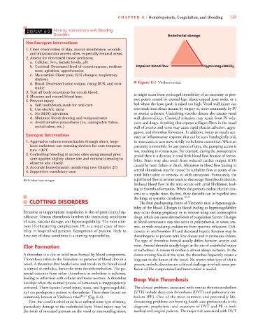
d. Renal: Decreased urine output, rising BUN, and crea- ■ Figure 6-4 Virchow’s triad.
tinine
3. Test all body secretions for occult blood. as might occur from prolonged immobility of an extremity or pres-
4. Measure and record blood loss.
5. Prevent injury. sure points caused by crossed legs, elastic-topped knee socks, or a
a. Soft toothbrush/swab for oral care bed where the knee gatch is raised too high. Vessel wall injury can
b. Use electric razor also result from direct trauma by surgery or, more commonly, by IV
c. No IM/SQ injections or arterial catheters. Underlying vascular disease also creates vessel
d. Minimize blood drawing and venipunctures wall abnormalities. Chemical irritation may result from IV solu-
e. Avoid invasive procedures (i.e., nasogastric tubes, tions and drugs. Anything that exposes collagen fibers in the vessel
rectal tubes, etc.) wall of arteries and veins may cause rapid platelet adhesion, aggre-
gation, and thrombus formation. In addition, injury to vessels acti-
Emergent Interventions
vates an inflammatory response that can be seen histologically and,
1. Aggressive volume resuscitation through short, large- in most cases, is seen most vividly in the lower extremities. When an
bore catheters; use warming devices for core tempera- extremity is immobile for any period of time, the pumping action is
ture 36 C lost, resulting in venous stasis. For example, during the postoperative
2. Controlling bleeding at access sites using manual pres- period there is a decrease in total limb blood flow because of immo-
sure applied slightly above site and minimal dressing to bility. Stasis may also result from reduced cardiac output (CO)
observe site closely
3. Accurate hemodynamic monitoring (see Chapter 21) caused by heart failure or shock. Alteration in blood flow leading to
4. Supportive ventilatory care arterial thrombosis may be caused by turbulent flow at points of ar-
terial bifurcation or stenosis, or with aneurysms. Fortunately, the
rapid blood flow in arteries tends to discourage thrombus formation.
BUN, blood urea nitrogen.
Reduced blood flow in the atria occurs with atrial fibrillation, lead-
ing to thrombus formation. When the patient’s cardiac rhythm con-
verts to a regular sinus rhythm, these thrombi can be expelled into
the lungs or systemic circulation. 16,17
CLOTTING DISORDERS The final predisposing factor of Virchow’s triad is hypercoagula-
bility of the blood. Changes in bloodleading to hypercoagulability
Excessive or inappropriate coagulation is also of great clinical sig- may occur during pregnancy or in women using oral contraceptive
nificance. Venous thrombosis involves the interacting conditions drugs, which can cause elevated levels of coagulation factors. Changes
of stasis, vascular damage, and hypercoagulability. The most com- in blood constituents may also occur in polycythemia, in severe ane-
mon life-threatening complication, PE, is a major cause of mor- mia, or with circulating endotoxins from systemic infections. Defi-
tality in hospitalized patients. Recognition of patients likely to ciencies in antithrombin III and decreased hepatic function may be
have any of these conditions is a nursing responsibility. thrombogenic in patients with liver disease and in premature infants.
The type of thrombus formed usually differs between arteries and
Clot Formation veins. Arterial thrombi usually begin at the site of endothelial injury
or turbulence. A venous thrombus is almost always occlusive. In the
A thrombus is a clot or solid mass formed by blood components. slower-moving blood of the veins, the thrombus frequently creates a
Thrombosis refers to the formation or presence of blood clots in a long cast in the lumen of the vessel. No matter what type of clot is
vessel. A thrombus that breaks loose and travels in the blood vessel present, embolic disorders are a clinical challenge in which tissue per-
is termed an embolus, hence the term thromboembolism. The po- fusion will be compromised and intervention is needed.
tential outcome from either thrombosis or embolism is ischemia,
leading to infarction with cellular and tissue necrosis. A thrombus Deep Vein Thrombosis
develops when the normal process of hemostasis is inappropriately
activated. Three factors (vessel injury, stasis, and hypercoagulabil- The clinical problems associated with venous thromboembolism
ity) can predispose a patient to thrombosis. These three factors are (VTE) include deep vein thrombosis (DVT) and pulmonary em-
commonly known as Virchow’s triad 16,17 (Fig. 6-4). bolism (PE). One of the most common and potentially life-
First, the vessel involved must have suffered some type of injury, threatening problems confronting health care professionals is the
particularly damage to the endothelial layer. Vessel injury may be diagnosis, prophylaxis, and treatment of DVT and PE in both
the result of sustained pressure on the vessel or surrounding tissue, medical and surgical patients. The major risk associated with DVT