
Page 292 - Cardiac Nursing
P. 292
8 P
M
8 P
1:1
1:1
M
g
g
Pa
Pa
Pa
/29
/29
6
xd
6
/09
1
1
1
/09
/09
a
a
ara
t
ara
a
c.
c.
In
a
In
68
68
e 2
g
e 2
A
p
t
p
A
p
xd
26
7-2
7-2
26
12_
0-c
K34
76.
LWB K34 0-c 12_ p p pp267-276.qxd 6/29/09 11:18 PM Page 268 Aptara Inc.
q
q
q
LWBK340-c12_
LWB
76.
268 P A R T III / Assessment of Heart Disease
viewed and interpreted. More recently, digital imaging technology
is used increasingly in radiology allowing for rapid viewing of CHEST FILM FINDINGS IN ACUTE
films on monitors rather than on light boxes. Computerized radi- CARE DETERMINING LINE, TUBE,
ographs can be viewed immediately on monitors on the CCU and AND CATHETER PLACEMENT
stored images allow the provider to readily compare current films
4
with previous images. To ensure that all anatomic structures are
seen, radiographs are read according to a certain pattern. This Bedside radiographs are used not only to assess for cardiopul-
method is called the directed search method. It is common prac- monary abnormalities, but also to evaluate placement oflines,
tice to look at soft tissues, bones, and diaphragms first, then at tubes, anddevices used in acute care. In addition to providing
the lungs from apex to base, and finally at the outline of the valuable information regarding the patient’s cardiopulmonary
heart and the aorta. Except for the heart, most structures in the status, the chest radiograph allows for early recognition of com-
chest are bilateral. Thus, if an abnormality is found on one side plications related to line placement as well as to evaluate thera-
of the chest, the other side should be observed to ensure that this peutic result after interventions such as drainage of a pleural ef-
“abnormality” is not present there. Even if an obvious abnor- fusion by chest tube placement. Table 12-1 lists invasive lines,
mality is present, a directed search should be completed so that tubes, anddevices commonly used in acute cardiovascular care
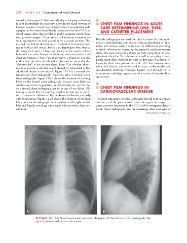
additional disease is not missed. Figure 12-1A is a normal pos- anddescribes radiologic findings. Figures 12-3 through 12-10
teroanterior chest radiograph; Figure 12-1B is a normal lateral demonstrate radiologic appearance of a variety of invasive lines
2
2
chest radiograph. Figure 12-2A shows the location of the lung and devices.
lobes on the frontal chest radiograph. Because some lobes are
anterior and some are posterior, an abnormality in a certain area
on a frontal chest radiograph can be in one of two lobes. Ob- CHEST FILM FINDINGS IN
taining a lateral film or noticing whether an anterior or poste- CARDIOVASCULAR DISEASE
rior structure is obliterated by an abnormal density can help
with localization. Figure 12-2B shows the location of the lung The chest radiograph provides useful data that aid in the complete
lobes on a lateral radiograph. Abnormalities of the right middle assessment of the patient with acute chest pain and suspected
lobe and lingula would go undetected with posterior chest aus- acute coronary syndrome in the CCU and/or emergency depart-
cultation. ment. Chest radiography aids in evaluating other etiologies of
(text continues on page 273)
A B
■ Figure 12-1 (A) Normal posteroanterior chest radiograph. (B) Normal lateral chest radiograph. The
spine is posterior and the heart is anterior.