
Page 296 - Cardiac Nursing
P. 296
g
g
Pa
g
72
72
e 2
e 2
Pa
1:1
8 P
1
1:1
M
Pa
8 P
M
a
a
a
a
c.
c.
In
In
ara
p
p
A
A
t
ara
p
t
1
7-2
7-2
26
26
q
q
76.
76.
LWBK340-c12_
K34
LWB K34 0-c 12_ pp267-276.qxd 6/29/09 11:18 PM Page 272 Aptara Inc.
LWB
p
p
0-c
12_
/29
/29
6
/09
/09
/09
q
1
xd
6
xd
272 P A R T III / Assessment of Heart Disease
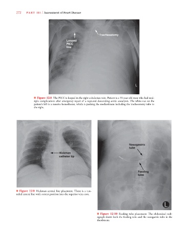
Tracheostomy
Looped
PICC
line
■ Figure 12-8 The PICC is looped in the right subclavian vein. Patient is a 55-year-old man who had mul-
tiple complications after emergency repair of a ruptured descending aortic aneurysm. The white-out on the
patient’s left is a massive hemothorax, which is pushing the mediastinum including the tracheostomy tube to
the right.
Nasogastric
tube
Hickman
catheter tip
Feeding
tube
■ Figure 12-9 Hickman central line placement. There is a tun-
neled central line with correct position into the superior vena cava.
■ Figure 12-10 Feeding tube placement. The abdominal radi-
ograph shows both the feeding tube and the nasogastric tube in the
duodenum.