
Page 295 - Cardiac Nursing
P. 295
g
g
Pa
Pa
e 2
71
g
e 2
Pa
1:1
1:1
1
1
M
M
8 P
8 P
71
a
a
a
a
c.
c.
In
In
ara
p
p
A
A
t
ara
p
t
1
76.
76.
7-2
7-2
q
xd
xd
q
q
26
LWBK340-c12_
K34
LWB K34 0-c 12_ pp267-276.qxd 6/29/09 11:18 PM Page 271 Aptara Inc.
LWB
0-c
p
26
12_
p
/29
/29
/09
/09
/09
6
6
C HAPTER 12 / Radiologic Examination of the Chest 271
Proximal end
fractured pacer
lead
Pacer
generator
Atrial pacer lead
Ventricular pacer lead
Distal tip fractured
pacer lead
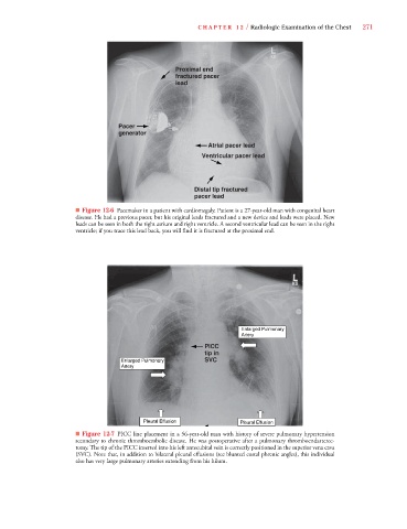
■ Figure 12-6 Pacemaker in a patient with cardiomegaly. Patient is a 27-year-old man with congenital heart
disease. He had a previous pacer, but his original leads fractured and a new device and leads were placed. New
leads can be seen in both the right atrium and right ventricle. A second ventricular lead can be seen in the right
ventricle; if you trace this lead back, you will find it is fractured at the proximal end.
Enlarged Pulmonary
Artery
PICC
tip in
Enlarged Pulmonary SVC
Artery
Pleural Effusion Pleural Effusion
■ Figure 12-7 PICC line placement in a 56-year-old man with history of severe pulmonary hypertension
secondary to chronic thromboembolic disease. He was postoperative after a pulmonary thromboendarterec-
tomy. The tip of the PICC inserted into his left antecubital vein is correctly positioned in the superior vena cava
(SVC). Note that, in addition to bilateral pleural effusions (see blunted costal phrenic angles), this individual
also has very large pulmonary arteries extending from his hilum.