Page 311 - Cardiac Nursing
P. 311

M
                                          M
                                        2 A
                                          M
                                              g
                                            Pa
                                            Pa
                                        2 A
                                  009
                                  009
                                6/2
                                     1
                                      0:4
                                      0:4
                                     1
                                              g
                                                    t
                                                   p
                                                   p
                                                    t
                                                     ara
                                                     ara
                                                     ara
                                                   p
                                               e 2
                                               e 2
                                              g
                                                87
                                                  A
                                                  A
                                                87
                    27
                    27
                      7-2
                        90.
                      7-2
                 13_
         LWBK340-c13_
         LWB K34 0-c 13_ p p pp277-290.qxd  30/06/2009  10:42 AM  Page 287 Aptara
         LWB
               0-c
            K34
                        90.
                              0/0
                              0/0
                             3
                              3
                             3
                                6/2
                          q
                          q
                          q
                           xd
                           xd
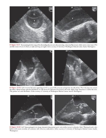
                   ■ Figure 13-17 Transesophageal echo image of the descending thoracic aorta demonstrating a dissection flap (arrow), which creates a true lumen (TL)w
                   and false lumen (FL) in the short axis (left) and long axis (right). (Echo courtesy of University of Washington Medical Center, Seattle, Washington.)


                   ■ Figure 13-18 Left: A normal left atrial appendage (LAA) is a pouch-like structure arising from the left atrium. This LAA does not contain
                   thrombus. Mitral stenosis and atrial fibrillation are risk factors for thrombus formation in the LAA. Right: Large thrombus (*) within the LAA.
                   This patient had atrial fibrillation. (Echo courtesy of University of Washington Medical Center, Seattle, Washington.)


                   ■ Figure 13-19 Left: Transesophageal echo image demonstrating normal aortic valve leaflets (arrow) in diastole. Right: Thickened aortic valve
                   leaflets with masses attached, all consistent with infectious endocarditis. (Echo courtesy of University of Washington Medical Center, Seattle,
                   Washington.)
   306   307   308   309   310   311   312   313   314   315   316