
Page 313 - Cardiac Nursing
P. 313
g
0/0
0/0
Pa
g
g
3
xd
q
q
3
3
xd
6/2
M
2 A
2 A
Pa
M
M
0:4
009
009
6/2
0:4
1
1
q
t
ara
ara
p
p
t
ara
LWB
LWBK340-c13_
LWB K34 0-c 13_ p p pp277-290.qxd 30/06/2009 10:42 AM Page 289 Aptara
13_
0-c
K34
7-2
27
27
90.
90.
7-2
e 2
A
A
p
89
e 2
89
C HAPTER 1 3 / Echocardiography 289
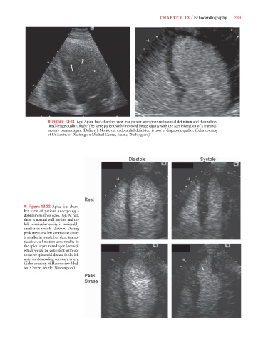
■ Figure 13-21 Left: Apical four-chamber view in a patient with poor endocardial definition and thus subop-
timal image quality. Right: The same patient with improved image quality with the administration of a transpul-
monary contrast agent (Definity). Notice the endocardial definition is now of diagnostic quality. (Echo courtesy
of University of Washington Medical Center, Seattle, Washington.)
■ Figure 13-22 Apical four-cham-
ber view of patient undergoing a
dobutamine stress echo. Top: At rest,
there is normal wall motion and the
left ventricular cavity is noticeably
smaller in systole. Bottom: During
peak stress, the left ventricular cavity
is smaller in systole but there is a no-
ticeable wall motion abnormality in
s
the apical septum and apex (arrows),
which would be consistent with ob-
structive epicardial disease in the left
anterior descending coronary artery.
(Echo courtesy of Harborview Med-
ical Center, Seattle, Washington.)