
Page 37 - Cardiac Nursing
P. 37
92806_c01.qxd 11/21/11 10:30 AM Page 13
CHAPTER 1 / Cardiac Anatomy and Physiology 13
Parasympathetic preganglionic cardiac nerves arise from the right
and left vagus nerves and synapse with postganglionic nerves close
to their target cardiac cells.
Both vagal and sympathetic cardiac nerves converge in the car-
diac plexus. The cardiac plexus is situated superior to the bifurca-
tion of the pulmonary artery, behind the aortic arch, and anterior
to the trachea at the level of tracheal bifurcation. From the cardiac
plexus, the cardiac nerves course in two coronary plexuses along
with the right and left coronary blood vessels.
Sympathetic fibers are richly distributed throughout the heart.
Right sympathetic ganglia fibers most commonly innervate the si-
nus node, the right atrium, the anterior ventricular walls, and to
some extent the AV node. Most commonly, left sympathetic gan-
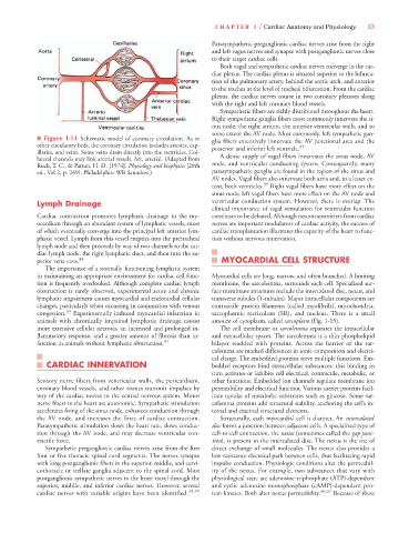
■ Figure 1-14 Schematic model of coronary circulation. As in glia fibers extensively innervate the AV junctional area and the
other circulatory beds, the coronary circulation includes arteries, cap- posterior and inferior left ventricle. 35
illaries, and veins. Some veins drain directly into the ventricles. Col-
lateral channels may link arterial vessels. Art, arterial. (Adapted from A dense supply of vagal fibers innervates the sinus node, AV
Ruch, T. C., & Patton, H. D. [1974]. Physiology and biophysics [20th node, and ventricular conducting system. Consequently, many
ed., Vol 2, p. 249]. Philadelphia: WB Saunders.) parasympathetic ganglia are found in the region of the sinus and
AV nodes. Vagal fibers also innervate both atria and, to a lesser ex-
tent, both ventricles. 35 Right vagal fibers have more effect on the
sinus node; left vagal fibers have more effect on the AV node and
Lymph Drainage ventricular conduction system. However, there is overlap. The
clinical importance of vagal stimulation for ventricular function
Cardiac contraction promotes lymphatic drainage in the my- continues to be debated. Although neurotransmitters from cardiac
ocardium through an abundant system of lymphatic vessels, most nerves are important modulators of cardiac activity, the success of
of which eventually converge into the principal left anterior lym- cardiac transplantation illustrates the capacity of the heart to func-
phatic vessel. Lymph from this vessel empties into the pretracheal tion without nervous innervation.
lymph node and then proceeds by way of two channels to the car-
diac lymph node, the right lymphatic duct, and then into the su-
perior vena cava. 34 MYOCARDIAL CELL STRUCTURE
The importance of a normally functioning lymphatic system
in maintaining an appropriate environment for cardiac cell func- Myocardial cells are long, narrow, and often branched. A limiting
tion is frequently overlooked. Although complete cardiac lymph membrane, the sarcolemma, surrounds each cell. Specialized sur-
obstruction is rarely observed, experimental acute and chronic face membrane structures include the intercalated disc, nexus, and
lymphatic impairment causes myocardial and endocardial cellular transverse tubules (T-tubules). Major intracellular components are
changes, particularly when occurring in conjunction with venous contractile protein filaments (called myofibrils), mitochondria,
congestion. 34 Experimentally induced myocardial infarction in sarcoplasmic recticulum (SR), and nucleus. There is a small
animals with chronically impaired lymphatic drainage causes amount of cytoplasm, called sarcoplasm (Fig. 1-15).
more extensive cellular necrosis, an increased and prolonged in- The cell membrane or sarcolemma separates the intracellular
flammatory response, and a greater amount of fibrosis than in- and extracellular spaces. The sarcolemma is a thin phospholipid
farction in animals without lymphatic obstruction. 34 bilayer studded with proteins. Across the barrier of the sar-
colemma are marked differences in ionic composition and electri-
cal charge. The embedded proteins serve multiple functions. Em-
CARDIAC INNERVATION bedded receptors bind extracellular substances; this binding in
turn activates or inhibits cell electrical, contractile, metabolic, or
Sensory nerve fibers from ventricular walls, the pericardium, other functions. Embedded ion channels regulate membrane ion
coronary blood vessels, and other tissues transmit impulses by permeability and electrical function. Various carrier proteins facil-
way of the cardiac nerves to the central nervous system. Motor itate uptake of metabolic substrates such as glucose. Some sar-
nerve fibers to the heart are autonomic. Sympathetic stimulation colemma proteins add structural stability, anchoring the cell’s in-
accelerates firing of the sinus node, enhances conduction through ternal and external structural elements.
the AV node, and increases the force of cardiac contraction. Structurally, each myocardial cell is distinct. An intercalated
Parasympathetic stimulation slows the heart rate, slows conduc- disc forms a junction between adjacent cells. A specialized type of
tion through the AV node, and may decrease ventricular con- cell-to-cell connection, the nexus (sometimes called the gap junc-
tractile force. tion), is present in the intercalated disc. The nexus is the site of
Sympathetic preganglionic cardiac nerves arise from the first direct exchange of small molecules. The nexus also provides a
four or five thoracic spinal cord segments. The nerves synapse low-resistance electrical path between cells, thus facilitating rapid
with long postganglionic fibers in the superior, middle, and cervi- impulse conduction. Physiologic conditions alter the permeabil-
cothoracic or stellate ganglia adjacent to the spinal cord. Most ity of the nexus. For example, two substances that vary with
postganglionic sympathetic nerves to the heart travel through the physiological state are adenosine triphosphate (ATP)-dependent
superior, middle, and inferior cardiac nerves. However, several and cyclic adenosine monophosphate (cAMP)-dependent pro-
cardiac nerves with variable origins have been identified. 33,35 tein kinases. Both alter nexus permeability. 36,37 Because of these