
Page 39 - Cardiac Nursing
P. 39
92806_c01.qxd 11/21/11 10:30 AM Page 15
CHAPTER 1 / Cardiac Anatomy and Physiology 15
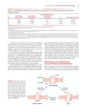
Table 1-2 ■ APPROXIMATE INTRACELLULAR AND EXTRACELLULAR ION CONCENTRATIONS AND ACTIVITIES IN
CARDIAC MUSCLE *
Ratio of Extracellular to
Extracellular Intracellular Intracellular
Ion † Concentration ‡ Concentration §11 Concentration E 1 Intracellular Activity #
Na 145 mM 15 mM 9.7 60 mV 7.0 mM
K 4 mM 150 mM 0.027 94 mV 125 mM
Cl 120 mM 5 mM 24 83 mV 15 mM
Ca 2 2 mM 10 4 M 2 10 4 129 mV 8 10 6 mM
* Values given are approximations and vary according to the cardiac tissue, species, and method used for measurement.
2
† Na , sodium; K , potassium; Cl , chloride; Ca , calcium.
–
‡ mM, millimolar.
§11 Most of the intracellular calcium is bound to proteins or sequestered in intracellular organelles; thus, total intracellular calcium content approximates 1 to 2 mm. During contrac-
tion, measurable intracellular calcium concentration approximates 10 –5 mm.
¶ E 1 , equilibrium potential; mV, millivolt.
# Median values from summarized data; these values should be considered as subject to revision. Concentrations and equilibrium potentials from Sperelakis, N. (1979). Origin of the
cardiac resting potential. In R. M. Berne (Ed.), Handbook of physiology, section 2: The cardiovascular system, vol 1, the heart (p. 193). Bethesda: American Physiological Society. Activi-
ties are approximations from Lee, C. O. (1981). Ionic activities in cardiac muscle cells and application of ion-sensitive microelectrodes, American Journal of Physiology, 10, H461,
H464 and Fozzard, H. A., & Wasserstrom, J. A. (1985). Voltage dependence of intracellular sodium and control of contraction. In P. P. Zipes, & J. Jalife. Cardiac electrophysiology
and arrhythmias (p. 52.). Orlando: Grune & Tratton.
For each ion, concentration differences across the sarcolemma layer or span its full thickness. Some of the proteins contain a water-
are determined by the sarcolemma’s permeability to that ion and filled pore that spans the membrane, connecting the intracellular
the balance of forces moving the ion from one to the other side of and extracellular spaces and forming a channel through which ions
the membrane. Electrical and concentration differences are main- can pass. Membrane channels open and close in response to a stim-
tained by a number of active and passive processes. Typical con- ulus (electrical, mechanical, or chemical), allowing passage of specific
centration differences are outlined in Table 1-2. ions when open. The opening and closing properties of a channel are
The sarcolemma is composed of phospholipid molecules. Each called its gating characteristics. The ability of a channel to selectively
molecule consists of a charged hydrophilic (water-attracting) glob- allow passage of certain ions while restricting other ions is called its
ular head and a noncharged hydrophobic (water-repelling) tail. selectivity property. Many ion channels are named after the ion for
The molecules organize into thin sheets, with the heads oriented in which they have selectivity. Some common types are sodium chan-
a consistent direction. Two sheets are aligned tail-to-tail to form a nels, potassium channels, and calcium channels (Fig. 1-16).
double layer (bilayer). The tails form the core of the sheet, and the
heads are directed outward in both directions. The result is a 7- to Mechanisms of Ion Distribution
9-nm, high-resistance, insulated barrier to ionic movement. across the Myocardial Membrane
Proteins embedded within the phospholipid bilayer may com-
pose more than half the mass of the membrane. Proteins function as Ions are distributed across the sarcolemma according to the mem-
receptors, channels, pumps, or structural stabilizers. The proteins brane permeability to the ion and the electrical and diffusion
may be inserted into the intracellular or extracellular side of the bi- forces on the ion. For each ion that can penetrate the membrane,
■ Figure 1-16 Three states of a
voltage-gated ion channel. Depicted
are the two closed and one open
state. Transition between these states
(arrows) open the channel (activa-
tion), close the channel in a refrac-
tory state where it cannot be re-
opened (inactivation), and reactivate
the channel by ending this refractory
state (recovery). (From Katz, A.
[2006]. Physiology of the heart [4th
ed., p. 376]. Philadelphia: Lippincott
Williams & Wilkins.)