
Page 38 - Cardiac Nursing
P. 38
92806_c01.qxd 11/21/11 10:30 AM Page 14
14 PA R T I / Anatomy and Physiology
orderly alignment of contractile proteins into myofilaments gives
the myocardial cell its striated (striped) appearance.
Mitochondria are small, rod-shaped membranous structures lo-
cated within the cell. Substrate breakdown and high-energy com-
pound synthesis occurs within the mitochondria. The relative
abundance of mitochondria in cardiac muscle cells reflects the
high level of biochemical activity required to support the heart’s
continuous contractile activity.
The SR is an extensive, self-contained internal membrane sys-
tem. The T-tubules and SR link membrane depolarization to the
mechanical activity of the contractile protein filaments. This func-
tional coordination is called excitation–contraction coupling. The
SR is the major storage depot for calcium ion, which releases then
takes up calcium ions with each contraction of the heart.
The nucleus contains the genetic material of the cell. The nu-
cleus is the site where new proteins are synthesized.
MYOCARDIAL CELL ELECTRICAL
CHARACTERISTICS
There is an electrical potential difference across the sarcolemma;
it is measured in millivolts (mV). During the interim between ex-
citations, the intracellular space is negative compared with the ex-
tracellular space. This potential difference is called the membrane
resting potential. During excitation, the potential difference
changes: the inside of the cell becomes less negative or slightly
positive compared with the extracellular space. This type of po-
tential difference change is called depolarization. After depolariza-
tion, the cell membrane repolarizes, or returns to the resting po-
tential value. The normal depolarization–repolarization cycle is
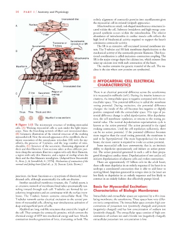
■ Figure 1-15 The microscopic structure of working myocardial known as the action potential. The action potential is the signal
cells. (A) Working myocardial cells as seen under the light micro- evoking contraction. Until the cell repolarizes sufficiently, there
scope. Note the branching network of fibers and intercalated discs. can be no action potential. If the potential difference becomes
(B) Schematic illustration of the internal structure of the working more negative than the usual resting potential, the membrane is
myocardial cell. Note the striated appearance of the myofibrils, the in-
timate association of the sarcoplasmic reticulum (SR) with the my- said to be hyperpolarized. The more hyperpolarized the mem-
ofibrils, the presence of T-tubules, and the large number of mito- brane, the more current is required to evoke an action potential.
chondria. (C) Structure of the sarcomere, illustrating alignment of Some myocardial cells have automaticity, that is, an intrinsic
thick and thin filaments. Cross sections taken at three different posi- ability to depolarize spontaneously and initiate an action poten-
tions along the sarcomere illustrate a region with only thick filaments, tial. The action potential generated in such a cell is then propa-
a region with only thin filaments, and a region of overlap where the gated throughout cardiac tissue. Depolarization of one cardiac cell
thick and the thin filaments interdigitate. (Adapted from Braunwald, initiates depolarization of adjacent cells and evokes contraction.
E., Ross, J., & Sonneblick, E. [1976]. Mechanisms of contraction of the There are approximately 19 billion cells in the adult heart;
normal and failing heart [2nd ed., p. 3]. Boston: Little, Brown.)
these cells must depolarize in an orderly sequence if the heart is to
undergo a coordinated contraction that is able to add force to
moving blood. Impulses generated in ectopic sites in the heart are
junctions, the heart functions as a syncytium of electrically coor- less likely to depolarize in an orderly sequence and less likely to
dinated cells, although anatomically the cells are discrete. contract in an orderly fashion that effectively pumps blood.
Another specialized membrane structure, the T-tubule system, is
an extensive network of membrane-lined tubes systematically tun- Basis for Myocardial Excitation:
neling inward through each cell. T-tubules are formed by sar- Characteristics of Biologic Membranes
colemma invaginations and are continuous with the surface mem-
brane. The T-tubule lumen contains extracellular fluid. The Intracellular and extracellular spaces are separated by a thin insu-
T-tubular network carries electrical excitation to the central por- lating membrane, the sarcolemma. These spaces have very differ-
tions of myocardial cells, allowing near-simultaneous activation of ent ionic compositions. The intracellular space contains high con-
deep and superficial parts of cells. centrations of potassium ion (positively charged) and protein
Myofibrils are long, rod-like structures that extend the length of (negatively charged) and has low concentration of sodium ion
the cell. They contain the contractile proteins, which convert the (positively charged). The extracellular space consists of high con-
chemical energy of ATP into mechanical energy and heat. Muscle centrations of sodium ion and chloride ion (negatively charged);
contraction involves generation of force, shortening, or both. The extracellular potassium ion concentration is low.