
Page 454 - Cardiac Nursing
P. 454
P
P
0-4
e 4
p42
8:2
009
M
g
7 A
qxd
0
38.
9/2
38.
30
30
Apt
9/0
ara
K34
0-c
19_
K34
L L LWB K34 0-c 19_ p42 0-4 38. qxd 0 9/0 9/2 009 0 0 8:2 7 A M P a a g e 4 30 Apt ara
LWB
LWBK340-c19_19_p420-438.qxd 09/09/2009 08:27 AM Page 430 Aptara
430 P A R T III / Assessment of Heart Disease
variation is most likely due to differences in how exercise-induced determination of the presence and strength of peripheral pulses
arrhythmias have been defined. Some studies have demonstrated should be made so that posttest comparisons are possible. Leg fatigue
that the occurrence of PVCs during an exercise test has minimal not related to claudication is often experienced at maximum exercise;
prognostic impact and should be interpreted in the context of “the a careful distinction should be made between these two symptoms.
company they keep,” 4,63 such that the decision to terminate the Dizziness and lightheadedness may reflect cerebral hypoxia and
test should be made on the basis of the patient’s history and may coincide with a feeling of exhaustion at maximum exercise.
whether the patient remains hemodynamically stable or the ar- Lightheadedness can also be a sign of left ventricular dysfunction
rhythmias are accompanied by symptoms. Other studies have or hypotension. Dizziness may be accompanied by signs of gray or
shown a clear association between PVCs that occur during exer- ashen pallor, diaphoresis, ataxic gait, dyspnea, and strained appear-
cise, recovery, or both, and increased mortality. 17,64–66 ance as blood is maximally shunted to the exercising muscles.
Trained observers should be able to recognize these responses and
Subjective Responses make a determination as to when the test should be stopped.
Assessment of symptoms and perception of effort during the exercise
test are important to maximize safety, and these subjective measures TEST TERMINATION
yield valuable diagnostic information. Obtaining careful assessments
of subjective measures during the exercise test requires thorough ex-
planations to ensure that the patient understands what is expected The usual goal of the exercise test in patients with known or sus-
and how to communicate these responses to those conducting the pected disease is to achieve a maximal level of exertion. This per-
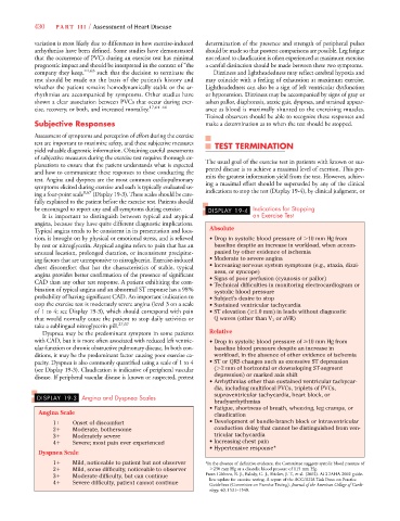
test. Angina and dyspnea are the most common cardiopulmonary mits the greatest information yield from the test. However, achiev-
symptoms elicited during exercise and each is typically evaluated us- ing a maximal effort should be superseded by any of the clinical
ing a four-point scale 8,67 (Display 19-3). These scales should be care- indications to stop the test (Display 19-4), by clinical judgment, or
fully explained to the patient before the exercise test. Patients should
be encouraged to report any and all symptoms during exercise. DISPLAY 19-4 Indications for Stopping
It is important to distinguish between typical and atypical an Exercise Test
angina, because they have quite different diagnostic implications.
Typical angina tends to be consistent in its presentation and loca- Absolute
tion, is brought on by physical or emotional stress, and is relieved • Drop in systolic blood pressure of 10 mm Hg from
by rest or nitroglycerin. Atypical angina refers to pain that has an baseline despite an increase in workload, when accom-
unusual location, prolonged duration, or inconsistent precipitat- panied by other evidence of ischemia
ing factors that are unresponsive to nitroglycerin. Exercise-induced • Moderate to severe angina
chest discomfort that has the characteristics of stable, typical • Increasing nervous system symptoms (e.g., ataxia, dizzi-
ness, or syncope)
angina provides better confirmation of the presence of significant • Signs of poor perfusion (cyanosis or pallor)
CAD than any other test response. A patient exhibiting the com- • Technical difficulties in monitoring electrocardiogram or
bination of typical angina and an abnormal ST response has a 98% systolic blood pressure
probability of having significant CAD. An important indication to • Subject’s desire to stop
stop the exercise test is moderately severe angina (level 3 on a scale • Sustained ventricular tachycardia
of 1 to 4; see Display 19-3), which should correspond with pain • ST elevation ( 1.0 mm) in leads without diagnostic
that would normally cause the patient to stop daily activities or Q waves (other than V 1 or aVR)
take a sublingual nitroglycerin pill. 27,67
Dyspnea may be the predominant symptom in some patients Relative
with CAD, but it is more often associated with reduced left ventric- • Drop in systolic blood pressure of 10 mm Hg from
ular function or chronic obstructive pulmonary disease. In both con- baseline blood pressure despite an increase in
ditions, it may be the predominant factor causing poor exercise ca- workload, in the absence of other evidence of ischemia
pacity. Dyspnea is also commonly quantified using a scale of 1 to 4 • ST or QRS changes such as excessive ST depression
(see Display 19-3). Claudication is indicative of peripheral vascular ( 2 mm of horizontal or downsloping ST-segment
disease. If peripheral vascular disease is known or suspected, pretest depression) or marked axis shift
• Arrhythmias other than sustained ventricular tachycar-
dia, including multifocal PVCs, triplets of PVCs,
supraventricular tachycardia, heart block, or
DISPLAY 19-3 Angina and Dyspnea Scales
bradyarrhythmias
• Fatigue, shortness of breath, wheezing, leg cramps, or
Angina Scale claudication
1
Onset of discomfort • Development of bundle-branch block or intraventricular
2
Moderate, bothersome conduction delay that cannot be distinguished from ven-
3
Moderately severe tricular tachycardia
4
Severe; most pain ever experienced • Increasing chest pain
• Hypertensive response*
Dyspnea Scale
1
Mild, noticeable to patient but not observer *In the absence of definitive evidence, the Committee suggests systolic blood pressure of
2
Mild, some difficulty, noticeable to observer 250 mm Hg or a diastolic blood pressure of 115 mm Hg.
3
Moderate difficulty, but can continue From Gibbons, R. J., Balady, G. J., Bricker, J. T., et al. (2002). ACC/AHA 2002 guide-
line update for exercise testing. A report of the ACC/AHA Task Force on Practice
4
Severe difficulty, patient cannot continue
Guidelines (Committee on Exercise Testing). Journal of the American College of Cardi-
ology,40, 1531–1540.