
Page 456 - Cardiac Nursing
P. 456
8:2
009
9/2
7 A
P
P
M
38.
0-4
p42
38.
9/0
0
qxd
K34
0-c
19_
K34
LWB K34 0-c 19_ p42 0-4 38. qxd 0 9/0 9/2 009 0 0 8:2 7 A M P a a g e 4 32 Apt ara
LWBK340-c19_19_p420-438.qxd 09/09/2009 08:27 AM Page 432 Aptara
L L LWB
32
32
g
e 4
ara
Apt
432 P A R T III / Assessment of Heart Disease
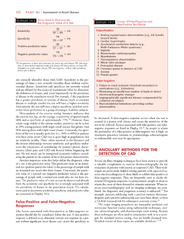
DISPLAY 19-6 Terms Used to Demonstrate Causes of False-Negative and
the Diagnostic Value of A Test DISPLAY 19-7
False-Positive Test Results
TP
Sensitivity 100 False-Positive
TP
FN
1. Resting repolarization abnormalities (e.g., left bundle-
TN
Specificity 100 branch block)
TN
FP 2. Cardiac hypertrophy
TP 3. Accelerated conduction defects (e.g.,
Positive predictive value 100
TP
FP Wolff–Parkinson–White syndrome)
4. Digitalis
TN
Negative predictive value 100 5. Nonischemic cardiomyopathy
TN
FN 6. Hypokalemia
7. Vasoregulatory abnormalities
TP, true-positives, or those with abnormal test results and with disease; FN, false-nega- 8. Mitral valve prolapse
tives, or those with normal test results with disease; FP, false-positives, or those with 9. Pericardial disease
abnormal test results and no disease; TN, true-negatives, or those with normal test 10. Coronary spasm in absence of CAD
results and no disease.
11. Anemia
12. Female gender
test correctly identifies those with CAD. Specificity is the per- False-Negative
centage of times a test correctly identifies those without cardio-
vascular disease. Sensitivity and specificity are inversely related 1. Failure to reach ischemic threshold secondary to
and are affected by the choice of discriminant value for abnormal, medications (e.g., -blockers)
the definition of disease, and, most importantly, by the prevalence 2. Monitoring an insufficient number of leads to detect
electrocardiographic changes
of disease in the population tested. For example, if the population 3. Angiographically significant disease compensated by
has a greater prevalence or severity of disease (such as coronary collateral circulation
disease in multiple vessels) the test will have a higher sensitivity. 4. Musculoskeletal limitations preceding cardiac
Alternatively, the test will have a higher specificity (and low sensi- abnormalities
tivity) when performed in a group of younger, healthier subjects.
Meta-analysis of the exercise testing literature indicates that
the exercise test has, on the average, a sensitivity of approximately be decreased. A false-negative response occurs when the test is
68% and a specificity of approximately 77%. 74 However, these normal in a person with disease and causes the sensitivity of the
values range widely in the various studies; sensitivity can be as low test to be reduced. Factors associated with false-positive and false-
as 40% among patients with single-vessel disease, but greater than negative responses are listed in Display 19-7. In people in whom
90% among those with triple-vessel disease. Conversely, the speci- the probability of a false-positive or false-negative test is high, an
ficity of the test is usually quite low (i.e., 50% to 60%) in patients alternative procedure (exercise or pharmacologic echocardiogram
who have more severe CAD but is quite high in populations that or radionuclide test) may be appropriate.
are relatively healthy. These values reported in the literature and
the inverse relationship between sensitivity and specificity under-
score the importance of considering the patient’s pretest charac-
teristics (chest pain and CAD risk factors) before beginning the ANCILLARY METHODS FOR THE
test. No test result can be interpreted accurately without consid- DETECTION OF CAD
ering the patient in the context of his or her pretest characteristics.
Another important term that helps define the diagnostic value Several ancillary imaging techniques have been shown to provide
of a test is the predictive value. The predictive value of an abnor- a valuable complement to exercise electrocardiography for the
mal test (positive predictive value) is the percentage of people with evaluation of patients with known or suspected CAD. These tech-
an abnormal test result who have disease. Conversely, the predic- niques are particularly helpful among patients with equivocal ex-
tive value of a normal test (negative predictive value) is the per- ercise electrocardiograms or those likely to exhibit false-positive or
centage of people with a normal test result who do not have dis- false-negative responses. They are frequently used to clarify ab-
ease. The predictive value of a test cannot be determined directly normal ST-segment responses in asymptomatic people or those in
from the sensitivity and specificity but is strongly associated with whom the cause of chest discomfort remains uncertain. When ex-
the prevalence of disease in the population tested. The calcula- ercise electrocardiography and an imaging technique are com-
tions used to determine sensitivity, specificity, and predictive value bined, the diagnostic and prognostic accuracy is enhanced. 75 For
are presented in Display 19-6. example, patients exhibiting both a positive exercise electrocar-
diogram and a positive radionuclide scan have been shown to have
False-Positive and False-Negative a 2.6-fold increased risk for subsequent coronary events. 76
Responses The major imaging procedures are myocardial perfusion and
ventricular function studies using radionuclide techniques, exer-
The factors associated with false-positive or false-negative re- cise echocardiography, and pharmacologic stress testing. Because
sponses should also be considered before the test. A false-positive these techniques are often used in conjunction with or as a surro-
response is defined as an abnormal exercise test response in a per- gate for standard exercise testing, they are briefly discussed here.
son without significant heart disease and causes the specificity to Detailed reviews of these topics are available elsewhere. 77,78