
Page 694 - Cardiac Nursing
P. 694
tar
Ap
670
AM
/1/
49
P
04.
qxd
7
5-7
g
p65
09
K34
LWBK340-c28_p655-704.qxd 7/1/09 9:9:49 AM Page 670 Aptara
28_
0-c
LWB
LWB K34 0-c 28_ p65 5-7 04. qxd 7 /1/ 09 9: 49 AM P a a g e e 670 Ap tar a a
670 PA R T I V / Pathophysiology and Management Disease
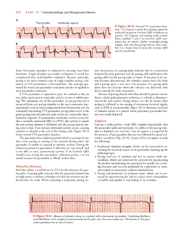
A
Pacing spike Ventricular capture
■ Figure 28-13 Normal VVI pacemaker func-
tion. (A) Capture is good, but sensing cannot be
evaluated because no intrinsic QRS complexes are
present. (B) Capture and sensing both normal.
Beats numbers 1 and 2 are intrinsic QRS com-
plexes that are sensed, inhibit ventricular pacing
B output, and reset the pacing interval. Beat num-
1 1 1 2 22 3 3 3 3 ber 3 is a fusion beat between the intrinsic QRS
and the paced beat.
heart. Pacemaker operation is evaluated by assessing these three tem, the presence of a pacing spike indicates that the connections
functions. Single-chamber pacemaker evaluation is much less between the pulse generator and the pacing cable and between the
complicated than dual-chamber evaluation. Because ventricular pacing cable and the pacing leads are intact. If any part of the sys-
pacing is the most common type of single-chamber pacing, eval- tem becomes disconnected, the stimulus cannot reach the body
uation of VVI pacemakers is discussed here. The concepts pre- and a pacing spike is not seen. The presence of a pacing spike
sented for ventricular pacemaker evaluation can also be applied to alone does not indicate where the stimulus was delivered, only
atrial pacemaker evaluation. that it entered the body somewhere.
A VVI pacemaker is expected to pace the ventricle at the set Absence of pacing stimuli when they should be present can in-
rate unless spontaneous ventricular activity occurs to inhibit pac- dicate a faulty pulse generator or battery, or a break or disconnec-
ing. The minimum rate of the pacemaker, or pacing interval, is tion in the lead system. Pacing stimuli can also be absent when
measured from one pacing stimulus to the next consecutive pac- pacing is inhibited by the sensing of extraneous electrical signals,
ing stimulus with no intervening sensed beats between the two. In such as EMI or myopotentials. Figure 28-14 illustrates total loss
a normally functioning VVI pacemaker, pacing spikes occur at the of stimulus release in a patient whose permanent pacemaker bat-
preset pacing interval and each spike results in a ventricular depo- tery was totally depleted.
larization (capture). If spontaneous ventricular activity occurs (ei-
ther a normally conducted QRS or a PVC), that activity is sensed, Capture
the next pacing stimulus is inhibited, and the pacing interval tim- Capture is indicated by a wide QRS complex immediately after
ing cycle is reset. If no intrinsic ventricular activity occurs, a pacing the pacemaker spike and represents the ability of the pacing stim-
stimulus is released at the end of the timing cycle. Figure 28-13 ulus to depolarize the ventricle. Loss of capture is recognized by
shows normal VVI pacemaker function. the presence of pacing spikes that are not followed by paced ven-
The pacemaker has a refractory period, which is a period of timed d tricular complexes (Fig. 28-15). Causes of loss of capture include
after either pacing or sensing in the ventricle during which the the following:
pacemaker is unable to respond to intrinsic activity. During the 1. Inadequate stimulus strength, which can be corrected by in-
refractory period, the pacemaker in effect has its “eyes closed” and creasing the electrical output of the pacemaker (turning up the
is not able to sense spontaneous activity. If an intrinsic QRS milliamperage).
should occur during the pacemaker’s refractory period, it is not 2. Pacing lead out of position and not in contact with my-
sensed because the pacemaker is “blind” at that time.
ocardium, which can sometimes be corrected by repositioning
the patient; repositioning the pacing lead is usually not a nurs-
Stimulus Release ing function and must be performed by a physician or some-
Stimulus release is verified on the ECG by the presence of a pac- one trained in intracardiac catheter manipulation.
ing spike. A pacing spike indicates that the pacemaker battery has 3. Pacing lead positioned in infarcted tissue, which can be cor-
enough power to initiate a stimulus and that the stimulus was de- rected by repositioning the lead to a place where myocardium
livered into the body. When evaluating a temporary pacing sys- is healthy and capable of responding to the stimulus.
■ Figure 28-14 Absence of stimulus release in a patient with a permanent pacemaker. Underlying rhythm is
atrial fibrillation with complete atrioventricular block and a very slow ventricular rate. The battery in the pace-
maker generator was at end of service.