
Page 699 - Cardiac Nursing
P. 699
AM
49
tar
P
7
09
/1/
5-7
04.
qxd
p65
Ap
675
g
K34
0-c
28_
LWB K34 0-c 28_ p65 5-7 04. qxd 7 /1/ 09 9: 49 AM P a a g e e 675 Ap tar a a
L L LWB
LWBK340-c28_p655-704.qxd 7/1/09 9:9:49 AM Page 675 Aptara
C HAPTER 2 8 / Pacemakers and Implantable Defibrillators 675
1 2 3 4 5 6 7 always easy to see. The atrial response to pacing is often so small
that it cannot be seen in many monitoring leads, so we cannot rely
on the presence of a P wave after atrial pacing spikes as evidence of
atrial capture. In the absence of a clear P wave, atrial capture can
be assumed only when an atrial pacing spike is followed by a nor-
mally conducted QRS complex within the programmed AV de-
lay. If the atrial spike captures the atrium and there is intact AV
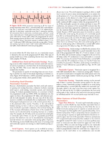
■ Figure 28-20 DDD pacemaker operating in all four states of conduction, the presence of the normal QRS indicates that the
pacing (stimulated strip). Beat 1, atrioventricular (AV) sequential pac- atrium must have been captured for conduction to have occurred
ing; beat 2, atrial pace, ventricular sense; beat 3, AV sequential pac- into the ventricles before the ventricular pacing stimulus was de-
ing; beat 4, atrial pace, ventricular sense; beat 5, premature ventricu- livered. Because a DDD pacemaker paces the ventricle at a preset
lar contraction; beat 6, atrial sense, ventricular pace; and beat 7, atrial AV delay after atrial pacing, the presence of a ventricular paced
sense, ventricular pace. Atrial capture is proven by beats 2 and 4 (atrial
spike followed by normal QRS within the programmed AV delay). beat after an atrial paced beat does not verify atrial capture, because
Atrial sensing is proven by beats 6 and 7 (normal P followed by paced the ventricle paces at the end of the AV delay regardless of whether
V at end of AV delay). Ventricular capture is verified by beats 1, 3, 6, atrial capture occurs. Therefore, atrial capture can be assumed only
and 7 (wide-paced QRS following ventricular pacing spike). Ventric- when there is an obvious P wave after every atrial pacing spike or
ular sensing is proven by beats 2 and 4 (atrial spike followed by nor- when an atrial pacing spike is followed by a normal QRS within
mal QRS, which inhibited ventricular pacing spike). the programmed AV delay (see Figs. 28-19B and 28-20).
Atrial Sensing. Atrial sensing is verified by the presence of an
intrinsic P wave that is followed by a paced ventricular beat at the
ity occurs before the AV delay times out, so a ventricular output end of the programmed AV delay. If a P wave is sensed, it starts
is released at the end of the programmed AV delay. This type of the AV delay and ventricular pacing is triggered at the end of the
pacing would occur if the underlying rhythm were sinus rhythm AV delay, unless AV conduction is intact and results in a normal
with complete AV block. QRS. The presence of a normal P wave followed by a normal QRS
proves only that AV conduction is intact, not that the P wave was
Inhibited State (Atrial and Ventricular Sensing). No pac-
sensed by the pacemaker. Therefore, atrial sensing is verified by an
ing occurs in either chamber because intrinsic atrial and ventricu-
intrinsic P wave followed by a paced QRS (see Figs. 28-19C and
lar activity is present at a rate faster than the minimum pacing rate
28-20).
(see Fig. 28-19D). This occurs when the underlying rhythm is
normal sinus rhythm. Ventricular Capture. Ventricular capture is recognized by a
The pacemaker is capable of switching from one state of pac- wide QRS immediately after a ventricular pacing spike. Ventricu-
ing to another on a beat-to-beat basis depending on intrinsic ac- lar capture is much easier to recognize than atrial capture and is the
tivity. Figure 28-20 illustrates a DDD pacemaker operating in all same as with single-chamber ventricular pacing (see Figs. 28-19A
four pacing states within a short period of time. and C and 28-20).
Ventricular Sensing. Ventricular sensing can be assessed
Evaluating Dual-Chamber only if there is intrinsic ventricular activity present for the pace-
Pacemaker Function maker to sense. Ventricular sensing is verified by an atrial pacing
Because a dual-chamber pacemaker has both atrial and ventricu- spike followed by a normal QRS that inhibits the ventricular pac-
lar pacing and sensing functions, evaluation includes assessing ing spike, which is the same event that proves atrial capture (see
atrial capture, atrial sensing, ventricular capture, and ventricular Figs. 28-19B and 28-20). If a QRS is sensed before the next atrial
sensing. To evaluate pacemaker function, it is necessary to know pacing spike is due, both the atrial and ventricular pacing stimuli
the programmed mode (e.g. DDD, DVI), the minimum rate, the are inhibited and the VA interval (atrial escape interval) is reset.
upper rate limit, the programmed AV delay, and refractory peri-
ods for both channels. In reality, the only time all of this infor- Other Features of Dual-Chamber
mation is available is immediately after an implantation, when the Pacemakers
final programmed parameters are in the current patient chart, or
in the physician’s office records. Therefore, in the real world of Upper-Rate Behavior. To avoid rapid ventricular pacing in
bedside nursing, we have to rely on a basic understanding of the response to atrial arrhythmias, dual-chamber pacemakers have an
issues involved in pacemaker evaluation, often without having all upper rate limit or maximal tracking rate that limits the rate at
of the necessary information at hand. Some of the needed infor- which ventricular pacing occurs in response to sensed atrial activ-
mation can be determined by measuring intervals on a rhythm ity. This upper rate limit applies only to paced tachycardias, not to
strip. For example, the AV delay can be measured from atrial spike intrinsic tachycardias. That is, tachycardias that are caused by ven-
to ventricular spike if there are any AV sequentially paced beats tricular pacing in response to rapid atrial rhythms should not ex-
present. The minimum rate can be determined by measuring the ceed the upper rate limit of the pacemaker. However, spontaneous
interval between two consecutive atrial pacing spikes, if present. VT or supraventricular tachycardia that conducts to the ventricle
The following sections briefly discuss the issues of assessing atrial through the normal AV node or across an accessory pathway may
and ventricular capture and sensing in a dual-chamber pacing sys- result in ventricular rates that exceed the upper rate limit of the
tem. pacemaker. When an atrial rate being tracked by the ventricular
channel of the pacemaker exceeds the upper rate limit, the pace-
Atrial Capture. Atrial capture can be verified by seeing a maker is programmed to limit the ventricular rate. Upper rate re-
P wave in response to every atrial pacing spike, although this is not sponses can be used alone or in combination and include