
Page 730 - Cardiac Nursing
P. 730
1
1
1
/09
/09
/09
4 A
M
M
2:0
2:0
4 A
/30
21.
q
q
5-7
5-7
21.
6
6
/30
q
xd
xd
Pa
t
ara
ara
p
p
t
In
c.
c.
a
a
In
p
g
g
e 7
Pa
Pa
g
06
A
A
e 7
06
06
70
K34
0-c
LWBK340-c29_
LWB K34 0-c 29_ pp705-721.qxd 6/30/09 12:04 AM Page 706 Aptara Inc.
LWB
29_
p
70
29_
p
706 PA R T I V / Pathophysiology and Management of Heart Disease
Table 29-1 ■ CLINICAL MANIFESTATIONS OF INFECTIVE
ENDOCARDITIS
Symptoms Physical Examination Findings
Fever Fever
Chills and sweats Changing or new heart murmur
Malaise Evidence of systemic emboli
Weight loss Splenomegaly
Anorexia Janeway lesions (small hemorrhages on palms or soles
Stroke symptoms of feet)
Myalgias Splinter hemorrhages (hemorrhagic streaks at finger
Arthralgias nail tips)
Confusion Osler’s nodules (small, tender nodules on finger or toe
CHF pads)
aggressive surgical intervention in cases complicated by CHF, inva-
sive abscesses, and prosthetic valve infections. 6
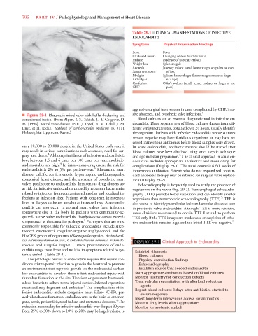
■ Figure 29-1 Rheumatic mitral valve with leaflet thickening and
commissural fusion. (From Alpert, J. S., Sabick, J., & Cosgrove, D. Blood cultures are an essential diagnostic tool in infective en-
M. [1998]. Mitral valve disease. In E. J. Topol, R. M. Califf, J. M. docarditis. Three separate sets of blood cultures drawn from dif-
Isner, et al. [Eds.], Textbook of cardiovascular medicine [p. 511]. ferent venipuncture sites, obtained over 24 hours, usually identify
Philadelphia: Lippincott-Raven.) the organism. Patients with infective endocarditis whose cultures
remain negative may have fastidious organisms or may have re-
ceived intravenous antibiotics before blood samples were drawn.
only 10,000 to 20,000 people in the United States each year, it In acute endocarditis, antibiotic therapy should be started after
may result in serious complications such as stroke, need for sur- blood cultures have been obtained using strict aseptic technique
3
gery, and death. Although incidence of infective endocarditis is and optimal skin preparation. The clinical approach in acute en-
2
low, between 1.5 and 6 cases per 100 cases per year, morbidity docarditis includes appropriate antibiotics and monitoring for
4
and mortality are high. In intravenous drug users, the risk for complications (Display 29-1). The usual course is 6 full weeks of
5
endocarditis is 2% to 5% per patient-year. Rheumatic heart intravenous antibiotics. Patients who do not respond well to stan-
disease, calcific aortic stenosis, hypertrophic cardiomyopathy, dard antibiotic therapy may be referred for surgical valve replace-
congenital heart disease, and the presence of prosthetic heart ment (Display 29-2).
valves predispose to endocarditis. Intravenous drug abusers are Echocardiography is frequently used to verify the presence of
at risk for infective endocarditis caused by recurrent bacteremias vegetations on the valves (Fig. 29-2). Transesophageal echocardio-
related to injection from contaminated needles and localized in- graphy (TEE) provides better resolution and can identify smaller
fections at injection sites. Patients with long-term intravenous vegetations than transthoracic echocardiography (TTE). TEE is
5
lines or dialysis catheters are also at increased risk. Acute endo- also useful to identify paravalvular leaks and annular abscesses seen
carditis can also occur in normal heart valves from infection in prosthetic valve endocarditis. Although TEE is more sensitive,
somewhere else in the body In patients with community-ac- some clinicians recommend to obtain TTE first and to perform
quired, native valve endocarditis, Staphylococcus aureus exceeds TEE only if the TTE images are inadequate or suspicion of infec-
5
streptococci as the causative pathogen. Pathogens that are most tive endocarditis remains high and the initial TTE was negative. 7
commonly responsible for subacute endocarditis include strep-
tococci, enterococci, coagulase-negative staphylococci, and the
HACEK group of organisms (Haemophilus species, Actinobacil-
lus actinomycetemcomitans, Cardiobacterium hominis, Eikenella DISPLAY 29-1 Clinical Approach to Endocarditis
species, and Kingella kingae). Clinical presentations of endo-
carditis range from fever and malaise to symptoms related to sys- Establish diagnosis
temic emboli (Table 29-1). Blood cultures
The pathologic process of endocarditis requires that several con- Physical examination findings
ditions exist to permit infection to grow in the heart and to promote Echocardiography
an environment that supports growth on the endocardial surface. Establish source that seeded endocarditis
For endocarditis to develop, there is first endocardial injury with Start appropriate antibiotics based on blood cultures
thrombus formation at the site. Transient or persistent bacteremia Monitor telemetry for conduction defects
allows bacteria to adhere to the injured surface. Infected vegetations Treat valvular regurgitation with afterload reduction
5
result and may fragment and embolize. The complications of in- agents
fective endocarditis include congestive heart failure (CHF), par- Repeat blood cultures 3 days after antibiotics started to
ensure response
avalvular abscess formation, embolic events to the brain or other or- Insert long-term intravenous access for antibiotics
4
gans, sepsis, pericarditis, renal failure, and metastatic abscesses. The Monitor drug levels when appropriate
reduction in mortality for infective endocarditis over the past 30 years Monitor for systemic emboli
from 25% to 30% down to 10% to 20% may be largely related to