Page 731 - Cardiac Nursing
P. 731

1
                                   2:0
                                   2:0
                                /09
                                    1
                                   1
                                        M
                                          Pa
                                          Pa
                                     4 A
                                     4 A
                                        M
                          q
                           xd
                           xd
                        21.
                          q
                          q
                              /30
                                /09
                                /09
                             6
                             6
                              /30
                                                  t
                                                  ara
                                                  ara
                                                 p
                                                 p
                                                  t
                                                     In
                                                      c.
                                                      c.
                                                   a
                                                   a
                                                     In
                                           g
                                            e 7
                                            e 7
                                          Pa
                                           g
                                           g
                                                A
                                                A
                                                 p
                                              07
                                              07
                                              07
                        21.
                   p
         LWB K34 0-c 29_ pp705-721.qxd  6/30/09  12:04 AM  Page 707 Aptara Inc.
         LWBK340-c29_
                    70
                    70
                   p
         LWB
                      5-7
                      5-7
                 29_
                 29_
               0-c
            K34
                                                                      C HAPTER 2 9 / Acquired Valvular Heart Disease  707
                    DISPLAY 29-2  Indications for Cardiac Surgery      DISPLAY 29-3 Risk Factors for Infective Endocarditis
                                 in Infective Endocarditis
                    Heart failure with hemodynamic instability          Recent dental procedure or periodontal disease
                    Persistent bacteremia and fever despite optimal antibiotic  History of congenital heart disease
                       therapy                                          History of valvular heart disease
                    Paravalvular abscess or fistula                      Long-term, in-dwelling intravenous line
                    Recurrence of endocarditis after full course of antibiotics  Genitourinary infections or instrumentation
                    Systemic emboli                                     Prosthetic valve (mechanical or biologic)
                    Heart failure due to prosthetic valvular dysfunction  History of intravenous drug abuse
                    Valve dehiscence (in prosthetic valvular endocarditis)  Hemodialysis
                    New conduction system defects
                    Fungal endocarditis
                     The American College of Cardiology/American Heart Associ-
                   ation (ACC/AHA) guidelines now recommend antibiotic prophy-  DIAGNOSTIC TESTING FOR
                   laxis for patients with prosthetic cardiac valves or rings; previous  VALVULAR HEART DISEASE
                   endocarditis; unrepaired cyanotic congenital heart disease; re-
                   paired congenital heart disease with prosthetic material or residual  The diagnosis of valvular heart disease is based on patient history,
                   defects adjacent to prosthetic device or patch; and cardiac trans-  physical assessment, anddiagnostic testing. Some tests, such as
                   plant recipients. 8
                                                                       the electrocardiogram and the chest radiograph, may be relatively
                                                                       insensitive in diagnosing valvular heart disease, even though they
                   Miscellaneous Causes of                             are part of the standard screening tests in patients withheart dys-
                   ValvularDisease                                     function. Both TTE and TEE are used to identify andquantify
                                                                       valvular heart disease. Diagnostic findings for specific valvular le-
                   Degenerative changes of the tissue, such as myxomatous degener-  sions are noted in the sections discussing each abnormality.
                   ation, calcification, and changes associated with Marfan syn-
                   drome, can cause valvular dysfunction. Trauma or infection may
                   affect the supportive or subvalvular apparatus. Dilation of the
                   ventricles caused by chronically elevated preloading may dilate an  MITRAL STENOSIS
                   atrioventricular valve opening to the point that the leaflets no
                   longer approximate and the valve becomes incompetent. Coro-  Cause
                   nary heart disease (CHD) and myocardial infarction can affect the  The predominant cause of mitral stenosis is rheumatic fever. The
                   papillary muscles of the right and left ventricles, causing either  mitral valve is the valve most often damagedby rheumatic cardi-
                   dysfunction caused by ischemia or frank flail of atrioventricular  tis. Rheumatic fever causes thickening anddecreased mobility of
                                                                         9
                   valve leaflets caused by papillary muscle rupture. Systemic diseases  the mitral valve leaflets associated withfusion of the commissures
                   such as lupus erythematosus and scleroderma may also cause  anddestruction of normalleaflet structure. Other conditions that
                   valvular dysfunction (see Display 29-3).            simulate the physiology of mitral stenosis include left atrial myx-
                                                                       oma, ball-valve left atrial thrombus, large left atrial endocarditis
                                                                       vegetations, or cor triatriatum (three atria). 10
                                                                       Pathology
                                                                       The rheumatic process causes the mitral valve to become fibri-
                                                                       nous, resulting in leaflet thickening, commissural or chordalfu-
                                                                       sion, and calcification. As a result, the mitral valve apparatus be-
                                                                       comes funnel shaped with a narrowed orifice. Fusion of the mitral
                                                                       valve commissures results in narrowing of the principal orifice,
                                                                       whereas interchordalfusion obliterates the secondary orifices.
                                                                       Pathophysiology
                                                                       Women have mitral stenosis more frequently than men. The nor-
                                                                                               2
                                                                       mal mitral valve area is 4 to 6 cm . Once the cross-sectional area
                                                                                                  2
                                                                       of the mitral valve is reduced to 2 cm or less, a pressure gradient
                                                                       between the left atrium andleft ventricle occurs. The reduced ori-
                                                                       fice impedes left atrial emptying. Increasedleft atrial pressure and
                                                                       dilation occurs along withleft atrialhypertrophy in an attempt to
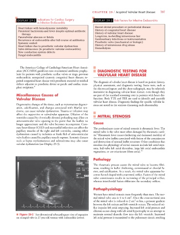
                   ■ Figure 29-2 Two-dimensional echocardiogram view of vegetation  maintain normaldiastolic flow into the left ventricle. Increased
                                                               w
                   on tricuspid valve in 27-year-old woman with endocarditis (arrow).  left atrial pressure is transmitted to the pulmonary circuit, resulting
   726   727   728   729   730   731   732   733   734   735   736