
Page 732 - Cardiac Nursing
P. 732
LWBK340-c29_p705-721.qxd 6/30/09 12:04 AM Page 708 Aptara Inc.
708 PA R T IV / Pathophysiology and Management of Heart Disease
in pulmonary hypertension and pulmonary congestion. Left atrial creased pulmonic S 2 intensity associated with pulmonary hyper-
enlargement may lead to atrial fibrillation and worsening of symp- tension (Table 29-2).
toms related to the loss of atrial kick. 10 Patients have left-sided Patients with mitral stenosis may exhibit malar blush (pink
CHF without left ventricular dysfunction. Mitral stenosis has a discoloration of the cheeks). Patients with severe mitral stenosis
sparing effect on the left ventricle. Symptoms of mitral stenosis may have weak pulses secondary to reduced cardiac output. The
are usually related to obstruction of the mitral valve rather than apical pulse is tapping in quality and is nondisplaced. A lower left
ventricular dysfunction. As pulmonary pressure increases, right- parasternal lift or heave caused by right ventricular hypertrophy
sided heart failure may occur. may be present. Cardiac rhythm is often irregularly irregular, in-
dicating atrial fibrillation.
Clinical Manifestations
Diagnostic Tests
Mild dyspnea on exertion occurs as the most common symptom
2
of mild mitral stenosis (valve area of 1.6 to 2.0 cm ). As mitral Echocardiography is used in the evaluation of mitral stenosis to (1)
2
stenosis becomes more severe (valve area of 1 to 1.5 cm ), dysp- quantify the valve area and gradient; (2) quantify the degree of mi-
nea, fatigue, paroxysmal nocturnal dyspnea, and atrial fibrillation tral insufficiency; (3) define the degree of left atrial enlargement;
may occur. When mitral stenosis becomes severe (valve area of (4) assess mitral annular calcification; (5) assess pulmonary artery
2
1 cm or less), symptoms include fatigue and dyspnea with mild pressures and degree of pulmonary hypertension; and (6) evaluate
exertion or rest. With advanced mitral stenosis, pulmonary hy- right- and left-sided ventricular function. A TEE provides better
pertension and symptoms of right-sided heart failure occur (i.e., detail of the mitral valve and better visualization of atrial throm-
edema, hepatomegaly, ascites, elevated jugular venous pressure). bus than does TTE. 12
Chest pain and hemoptysis may also occur. Increased left atrial Cardiac catheterization is used less in diagnosis of mitral steno-
pressure, atrial fibrillation, and stagnation of left atrial blood flow sis as echocardiography techniques improve. Cardiac catheteriza-
can result in the formation of mural thrombi, with resultant em- tion does allow for accurate assessment of valve area and can also
bolic events, including cerebral vascular accidents. Women who identify associated mitral regurgitation. For patients with known
had previously been asymptomatic with mitral stenosis may be- or suspected CHD, coronary angiography can delineate coronary
come symptomatic and even experience severe hemodynamic de- anatomy. Right heart catheterization can evaluate right heart and
compensation during pregnancy due to increased cardiac output pulmonary artery pressures.
and increased heart rate. Tachycardia reduces diastolic filling time Electrocardiography is nonspecific and does not indicate the
and worsens the mitral valve gradient while atrial fibrillation may severity of mitral stenosis. If the patient remains in sinus rhythm
precipitate pulmonary edema. 11 and left atrial enlargement has occurred, characteristic P mitrale
(broad, bifid P waves in leads II and V 1 ) may be identified. Right
Physical Assessment axis deviation and right ventricular hypertrophy may be noted in
severe mitral stenosis. Atrial fibrillation is common in patients with
In severe mitral stenosis, on auscultation, there are four typical long-standing mitral stenosis and is usually coarse in appearance.
findings including (1) an accentuated S 1 ; (2) an opening diastolic Chest radiography correlates with the degree of mitral stenosis.
snap; (3) a middiastolic rumble noted best at the apex (in sinus As mitral stenosis becomes more severe, the chest radiograph
rhythm), followed by presystolic accentuation; and (4) an in- demonstrates straightening of the left heart border caused by left
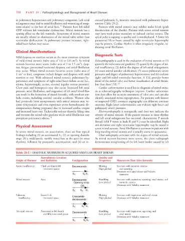
Table 29-2 ■ DIASTOLIC MURMURS IN ACQUIRED VALVULAR HEART DISEASE
Auscultatory Location Quality and
Origin of Murmur and Radiation Configuration Frequency Maneuvers That Alter Intensity
Aortic insufficiency Third and fourth left Blowing Increases with isometric exercise
intercostal spaces High pitched and squatting
Decreases with amyl nitrate and Valsalva
S 1 S 2 S 1 maneuver
Mitral stenosis Apex Rumbling Increases with expiration, squatting, amyl nitrate, and
Opening snap Low pitched isometric exercise
OS Decreases with Valsalva maneuver
S 1 S 2 S 1
Pulmonic Second left Crescendo- Blowing Increases with inspiration and amyl nitrate
insufficiency intercostal space High pitched Decreases with Valsalva maneuver
decrescendo
S 1 S 2 S 1
Tricuspid stenosis Parasternal at left fourth Decrescendo Rumbling Increases with inspiration, squatting, and
and fifth intercostal spaces Low pitched amyl nitrate
Decreases with Valsalva maneuver
S 1 S 2 S 1