
Page 230 - ACCCN's Critical Care Nursing
P. 230
Cardiovascular Assessment and Monitoring 207
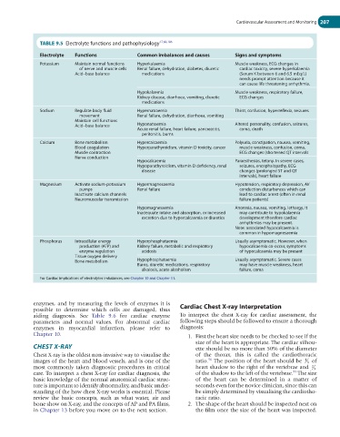
TABLE 9.5 Electrolyte functions and pathophysiology 17,66,106
Electrolyte Functions Common imbalances and causes Signs and symptoms
Potassium Maintain normal functions Hyperkalaemia Muscle weakness, ECG changes in
of nerve and muscle cells Renal failure, dehydration, diabetes, diuretic cardiac toxicity, severe hyperkalaemia
Acid–base balance medications (Serum K between 6 and 6.5 mEq/L)
needs prompt attention because it
can cause life threatening arrhythmia.
Hypokalaemia Muscle weakness, respiratory failure,
Kidney disease, diarrhoea, vomiting, diuretic ECG changes
medications
Sodium Regulate body fluid Hypernatraemia Thirst, confusion, hyperreflexia, seizures
movement Renal failure, dehydration, diarrhoea, vomiting
Maintain cell functions
Acid–base balance Hyponatraemia Altered personality, confusion, seizures,
Acute renal failure, heart failure, pancreatitis, coma, death
peritonitis, burns
Calcium Bone metabolism Hypercalcaemia Polyuria, constipation, nausea, vomiting,
Blood coagulation Hyperparathyroidism, vitamin D toxicity, cancer muscle weakness, confusion, coma,
Muscle contraction ECG changes (shortened QT intervals
Nerve conduction
Hypocalcaemia Paraesthesias, tetany. In severe cases,
Hypoparathyroidism, vitamin D deficiency, renal seizures, encephalopathy, ECG
disease changes (prolonged ST and QT
intervals), heart failure
Magnesium Activate sodium-potassium Hypermagnesaemia Hypotension, respiratory depression, AV
pumps Renal failure conduction disturbances which can
Inactivate calcium channels lead to cardiac arrest (often in renal
Neuromuscular transmission failure patients)
Hypomagnesaemia Anorexia, nausea, vomiting, lethargy, It
Inadequate intake and absorption, or increased may contribute to hypokalaemia
excretion due to hypercalcaemia or diuretics development therefore cardiac
arrhythmias may be present.
Note: associated hypocalcaemia is
common in hypomagnesaemia
Phosphorus Intracellular energy Hyperphosphataemia Usually asymptomatic. However, when
production (ATP) and Kidney failure, metabolic and respiratory hypocalcaemia co-occur, symptoms
enzyme regulation acidosis of hypocalcaemia may be present
Tissue oxygen delivery
Bone metabolism Hypophosphataemia Usually asymptomatic. Severe cases
Burns, diuretic medications, respiratory may have muscle weakness, heart
alkalosis, acute alcoholism failure, coma
For Cardiac implications of electrolytes imbalances, see Chapter 10 and Chapter 11.
enzymes, and by measuring the levels of enzymes it is Cardiac Chest X-ray Interpretation
possible to determine which cells are damaged, thus
aiding diagnosis. See Table 9.6 for cardiac enzyme To interpret the chest X-ray for cardiac assessment, the
parameters and normal values. For abnormal cardiac following steps should be followed to ensure a thorough
enzymes in myocardial infarction, please refer to diagnosis:
Chapter 10. 1. First the heart size needs to be checked to see if the
size of the heart is appropriate. The cardiac silhou-
CHEST X-RAY ette should be no more than 50% of the diameter
Chest X-ray is the oldest non-invasive way to visualise the of the thorax, this is called the cardiothoracic
96
images of the heart and blood vessels, and is one of the ratio. The position of the heart should be 1 3 of
most commonly taken diagnostic procedures in critical heart shadow to the right of the vertebrae and 2 3
93
care. To interpret a chest X-ray for cardiac diagnosis, the of the shadow to the left of the vertebrae. The size
basic knowledge of the normal anatomical cardiac struc- of the heart can be determined in a matter of
ture is important to identify abnormality, and basic under- seconds even for the novice clinician, since this can
standing of the how chest X-ray works is essential. Please be simply determined by visualising the cardiotho-
review the basic concepts, such as what water, air and racic ratio.
bone show on X-ray, and the concepts of AP and PA films, 2. The shape of the heart should be inspected next on
in Chapter 13 before you move on to the next section. the film once the size of the heart was inspected.