
Page 225 - ACCCN's Critical Care Nursing
P. 225
202 P R I N C I P L E S A N D P R A C T I C E O F C R I T I C A L C A R E
Flow-directed
catheter
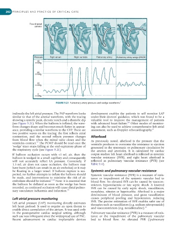
Pressure Right atrium Right ventricle Pulmonary artery Pulmonary artery wedge (PAOP)
30
mmHg
20
mmHg
10
mmHg
0
mmHg
FIGURE 9.21 Pulmonary artery pressure and wedge waveforms. 5
indirectly the left atrial pressure. The PAP waveform looks development enables the patients to self monitor LAP
similar to that of the arterial waveform, with the tracing under their doctors’ guidance, which was found to be a
showing a systolic peak, dicrotic notch and a diastolic dip valuable tool to improve the management of patients
(see Figure 9.21). When the balloon is inflated, the wave- with advanced heart failure. Other modes of monitor-
60
form changes shape and becomes much flatter in appear- ing can also be used to achieve comprehensive left atrial
ance, providing a similar waveform to the CVP. There are assessment, such as Doppler echocardiography. 61
two positive waves on the tracing: the first reflects atrial
contraction, and the second reflects pressure changes Afterload
from blood flow when the mitral valve closes and the As previously noted, afterload is the pressure that the
ventricles contract. The PCWP should be read once the ventricle produces to overcome the resistance to ejection
57
‘wedge’ trace stops falling at the end-expiratory phase of generated in the systematic or pulmonary circulation by
the respiratory cycle (see Figure 9.21). the arteries and arterioles. It is calculated by cardiac
If balloon occlusion occurs with <1 mL air, then the output studies: left heart afterload is reflected as systemic
balloon is wedged in a small capillary and consequently vascular resistance (SVR), and right heart afterload is
will not accurately reflect LA pressure. Conversely, if reflected as pulmonary vascular resistance (PVR) (see
1.5 mL air does not cause occlusion, the balloon may Table 9.4).
have burst (which can result in an air embolus) or it may
be floating in a larger vessel. If balloon rupture is sus- Systemic and pulmonary vascular resistance
pected, no further attempts to inflate the balloon should Systemic vascular resistance (SVR) is a measure of resis-
be made, and interventions to minimise the risk of air tance or impediment of the systemic vascular bed to
embolism should be initiated. 7,58 Note: it is essential that blood flow. An elevated SVR can be caused by vasocon-
the balloon be deflated as soon as the wedge has been strictors, hypovolaemia or late septic shock. A lowered
recorded, as continued occlusion will cause distal pulmo- SVR can be caused by early septic shock, vasodilators,
nary vasculature ischaemia and infarction. 59 morphine, nitrates or hypercarbia. Afterload is a major
determinant of blood pressure, and gross vasodilation
Left atrial pressure monitoring causes peripheral pooling and hypotension, reducing
SVR. The precise estimation of SVR enables safer use of
Left atrial pressure (LAP) monitoring directly estimates therapies such as vasodilators (e.g. sodium nitroprusside)
left heart preload. It used to require an open thorax to and vasoconstrictors (e.g. noradrenaline). 62
enable direct cannulation of the atrium. It was used only
in the postoperative cardiac surgical setting, although Pulmonary vascular resistance (PVR) is a measure of resis-
such use was infrequent since the widespread use of PAC. tance or the impediment of the pulmonary vascular
Recent advancement in cardiac implantable devices bed to blood flow. An elevated PVR (‘pulmonary