
Page 231 - ACCCN's Critical Care Nursing
P. 231
208 P R I N C I P L E S A N D P R A C T I C E O F C R I T I C A L C A R E
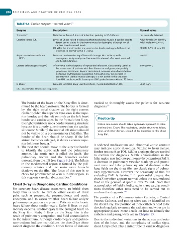
TABLE 9.6 Cardiac enzymes – normal values 91
Enzyme Description Normal value
Troponin T Detected within 4–6 hours of infarction, peaking in 10–24 hours. not normally detected
Creatine kinase (CK) Levels of CK are raised in diseases affecting skeletal muscle. It can be used to Adult female: 30–180 U/L
detect carrier status for Duchenne muscular dystrophy, although not all Adult male: 60–220 U/L
carriers have increased levels.
CK-MB is the first of cardiac enzymes to rise, levels peaking in 24 hours but CK-MB: 0–5% of total CK
returning to normal within 2–3 days.
Aspartate aminotransferase Detection and monitoring of liver cell damage. No cardiac-specific <40 U/L
(AST) isoenzymes; today rarely used because it is released after renal, cerebral
and hepatic damage.
Lactate dehydrogenase (LDH) Of no value in the diagnosis of myocardial infarction. Occasionally useful in 110–230 U/L
the assessment of patients with liver disease or malignancy (especially
lymphoma, seminoma, hepatic metastases); anaemia when haemolysis or
ineffective erythropoiesis suspected. Although it may be elevated in
patients with skeletal muscle damage, it is not useful in this situation.
Post-AMI, cardiac-specific isoenzyme LDH peaks between 48 and 72 hours.
1
D-Dimer Presence indicates deep vein thrombosis, myocardial infarction, DIC <0.25 ng/L
DIC = disseminated intravascular coagulation.
The border of the heart on the X-ray film is deter- needed to thoroughly assess the patients for accurate
mined by the heart anatomy. The border is formed diagnosis. 98
by: the right atrial shadow as the right convex
cardiac border; the superior vena cava as the supe-
rior border; and the left ventricle as the left heart
border and cardiac apex. In the frontal chest X-ray, Practice tip
the right ventricle is not a border-forming structure Critical care nurses should take a systematic approach to inter-
because it is directly superimposed on the cardiac preting chest X-rays. The respiratory, cardiac structures, tubes,
silhouette. Similarly, the normal left atrium should wires and other devices should all be identified in the chest
not be visible on a posteroanterior (PA) film. The X-ray film.
border of the heart should be sharp. If the left
atrium becomes enlarged, it shows a convex supe-
rior left heart border. 96 A widened mediastinum and abnormal aortic contour
3. The next step should move to the superior border may indicate aortic dissection. Similar to heart failure,
to identify the aortic arch and the pulmonary further tests such as TOE, MRI or angiography are needed
arteries. The aortic arch is called the knob. The to confirm the diagnosis. Subtle abnormalities in the
pulmonary arteries and the branches radiate hilar region may indicate pulmonary hypertension (PAH).
outward from the hili (see Figure 9.24). The hilum A decrease in pulmonary vascular markings and promi-
in the mediasternal region is formed by the pul- nent main and hilar pulmonary arterial shadows in the
monary arteries and the main stem bronchi lung fields on the chest film are classic signs of pulmo-
shadows on the film. The focus of this step is to nary hypertension. However the sensitivity of this for
check for prominence of vessels in this region, as excluding PAH is lacking. In pericardial disease, the
99
this suggests vascular abnormalities. 97
chest X-ray often appears normal unless the accumulated
fluid in the pericardial space is over 250 mL. Note that
Chest X-ray in Diagnosing Cardiac Conditions accumulation of fluid is indicated in many cardiac condi-
For coronary heart disease assessment, an initial chest tions therefore other tests need to be carried out to
X-ray film is useful to exclude other causes of chest confirm the diagnosis. 100
pain, such as pneumonia, pneumothorax and aortic
aneurysm, and to assess whether heart failure and/or The position of a Pulmonary Artery Catheter, a Central
pulmonary congestion are present. Patients with chronic Venous Catheter, and pacing wires can be identified on
heart failure show cardiomegaly, Kerby B lines or pul- the chest X-ray. The position of these catheters need to be
monary oedema. Cardiomegaly is the enlarged heart on checked regularly to ensure the catheters and wires are in
the X-ray film. Kerby B lines on the X-ray film is the appropriate places. More details on how to identify the
result of pulmonary congestion and fluid accumulation catheters and pacing wires are in Chapter 13.
in the interstitium. Although cardiomegaly and pulmo- Due to the individual variations in shape, size and rota-
nary oedema indicate heart failure, the chest X-ray alone tion of the heart, and the complexity of cardiac signs,
cannot diagnose the condition. Other forms of tests are chest X-rays often play a minor role in cardiac diagnosis.