
Page 228 - ACCCN's Critical Care Nursing
P. 228
Cardiovascular Assessment and Monitoring 205
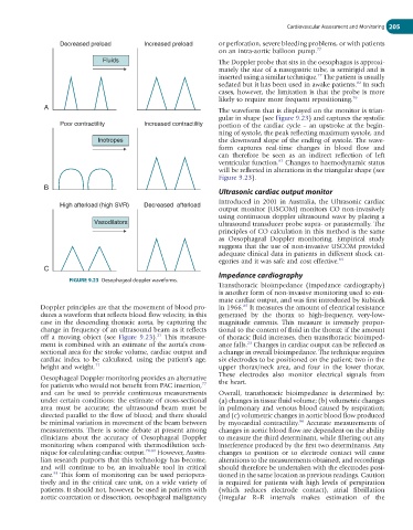
Decreased preload Increased preload or perforation, severe bleeding problems, or with patients
on an intra-aortic balloon pump. 77
Fluids The Doppler probe that sits in the oesophagus is approxi-
mately the size of a nasogastric tube, is semirigid and is
77
inserted using a similar technique. The patient is usually
82
sedated but it has been used in awake patients. In such
cases, however, the limitation is that the probe is more
likely to require more frequent repositioning. 76
A The waveform that is displayed on the monitor is trian-
gular in shape (see Figure 9.23) and captures the systolic
Poor contractility Increased contractility portion of the cardiac cycle – an upstroke at the begin-
ning of systole, the peak reflecting maximum systole, and
Inotropes the downward slope of the ending of systole. The wave-
form captures real-time changes in blood flow and
can therefore be seen as an indirect reflection of left
83
ventricular function. Changes to haemodynamic status
will be reflected in alterations in the triangular shape (see
Figure 9.23).
B
Ultrasonic cardiac output monitor
Introduced in 2001 in Australia, the Ultrasonic cardiac
High afterload (high SVR) Decreased afterload
output monitor (USCOM) monitors CO non-invasively
using continuous doppler ultrasound wave by placing a
Vasodilators ultrasound transducer probe supra- or parasternally. The
principles of CO calculation in this method is the same
as Oesophageal Doppler monitoring. Empirical study
suggests that the use of non-invasive USCOM provided
adequate clinical data in patients in different shock cat-
egories and it was safe and cost effective. 84
C
Impedance cardiography
FIGURE 9.23 Oesophageal doppler waveforms.
Transthoracic bioimpedance (impedance cardiography)
is another form of non-invasive monitoring used to esti-
mate cardiac output, and was first introduced by Kubicek
85
Doppler principles are that the movement of blood pro- in 1966. It measures the amount of electrical resistance
duces a waveform that reflects blood flow velocity, in this generated by the thorax to high-frequency, very-low-
case in the descending thoracic aorta, by capturing the magnitude currents. This measure is inversely propor-
change in frequency of an ultrasound beam as it reflects tional to the content of fluid in the thorax: if the amount
23
off a moving object (see Figure 9.23). This measure- of thoracic fluid increases, then transthoracic bioimped-
ment is combined with an estimate of the aorta’s cross- ance falls. Changes in cardiac output can be reflected as
23
sectional area for the stroke volume, cardiac output and a change in overall bioimpedance. The technique requires
cardiac index to be calculated, using the patient’s age, six electrodes to be positioned on the patient: two in the
height and weight. 77 upper thorax/neck area, and four in the lower thorax.
These electrodes also monitor electrical signals from
Oesophageal Doppler monitoring provides an alternative
77
for patients who would not benefit from PAC insertion, the heart.
and can be used to provide continuous measurements Overall, transthoracic bioimpedance is determined by:
under certain conditions: the estimate of cross-sectional (a) changes in tissue fluid volume; (b) volumetric changes
area must be accurate; the ultrasound beam must be in pulmonary and venous blood caused by respiration;
directed parallel to the flow of blood; and there should and (c) volumetric changes in aortic blood flow produced
be minimal variation in movement of the beam between by myocardial contractility. Accurate measurements of
86
measurements. There is some debate at present among changes in aortic blood flow are dependent on the ability
clinicians about the accuracy of Oesophageal Doppler to measure the third determinant, while filtering out any
monitoring when compared with thermodilution tech- interference produced by the first two determinants. Any
nique for calculating cardiac output. 78-80 However, Austra- changes to position or to electrode contact will cause
lian research purports that this technology has become, alterations to the measurements obtained, and recordings
and will continue to be, an invaluable tool in critical should therefore be undertaken with the electrodes posi-
81
care. This form of monitoring can be used periopera- tioned in the same location as previous readings. Caution
tively and in the critical care unit, on a wide variety of is required for patients with high levels of perspiration
patients. It should not, however, be used in patients with (which reduces electrode contact), atrial fibrillation
aortic coarctation or dissection, oesophageal malignancy (irregular R–R intervals makes estimation of the