
Page 277 - ACCCN's Critical Care Nursing
P. 277
254 P R I N C I P L E S A N D P R A C T I C E O F C R I T I C A L C A R E
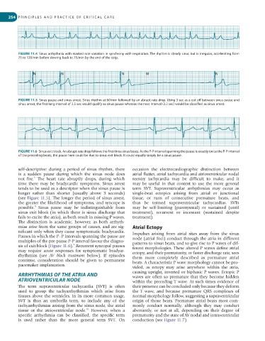
FIGURE 11.4 Sinus arrhythmia with marked rate variation in synchrony with respiration. The rhythm is clearly sinus but is irregular, accelerating from
75 to 120/min before slowing back to 75/min by the end of the strip.
N N N M N
FIGURE 11.5 Sinus pause and sinus arrest. Sinus rhythm at 60/min followed by an abrupt rate drop. Using 3 sec as a cut off between sinus pause and
sinus arrest, the first long interval of 2.5 sec would qualify as sinus pause whereas the next interval (3.2 sec) would be classified as sinus arrest.
FIGURE 11.6 Sinus exit block. An abrupt rate drop follows the first three sinus beats. As the P-P interval spanning the pause is exactly twice the P-P interval
of the preceding beats, the pause here could be due to sinus exit block. It could equally simply be a sinus pause.
self-descriptive: during a period of sinus rhythm, there occasion the electrocardiographic distinction between
is a sudden pause during which the sinus node does atrial flutter, atrial tachycardia and atrioventricular nodal
9
not fire. The heart rate abruptly drops, during which reentry tachycardia may be difficult to make, and it
time there may be bradycardic symptoms. Sinus arrest may be useful in that context to use the more general
tends to be used as a descriptor when the sinus pause is term SVT. Supraventricular arrhythmias may occur as
longer rather than shorter (usually above 3 seconds) single-beat ectopics arising from atrial or junctional
(see Figure 11.5). The longer the period of sinus arrest, tissue, or runs of consecutive premature beats, and
the greater the likelihood of symptoms, and syncope is thus be termed supraventricular tachycardias. SVTs
9
possible. Sinus pause may be indistinguishable from may be self-limiting (paroxysmal) or sustained (until
sinus exit block (in which there is sinus discharge that treatment), recurrent or incessant (sustained despite
fails to excite the atria), as both result in missing P waves. treatment).
The distinction is academic, however, as both arrhyth-
mias arise from the same groups of causes, and are sig- Atrial Ectopy
nificant only when they cause symptomatic bradycardia. Impulses arising from atrial sites away from the sinus
Pauses in which the P–P intervals spanning the pause are node (atrial foci) conduct through the atria in different
multiples of the pre-pause P-P interval favour the diagno- patterns to sinus beats, and so give rise to P waves of dif-
5
sis of exit block (Figure 11.6). Recurrent syncopal pauses ferent morphologies. These altered P waves define atrial
may require acute responses for symptomatic bradyar- ectopy, and their prematurity, or faster discharge rate, sees
rhythmias (see AV block treatment below). If episodes them more completely described as premature atrial
continue, consideration should be given to permanent beats. A characteristic P wave morphology cannot be pro-
pacemaker implantation.
vided, as ectopy may arise anywhere within the atria,
ARRHYTHMIAS OF THE ATRIA AND causing upright, inverted or biphasic P waves. Ectopic P
waves are often so premature that they become hidden
ATRIOVENTRICULAR NODE within the preceding T wave. At such times evidence of
The term supraventricular tachycardia (SVT) is often their presence can be concluded only because they deform
used to group the tachyarrhythmias which arise from the T wave, and because premature QRS complexes of
tissues above the ventricles. In its more common usage, normal morphology follow, suggesting a supraventricular
SVT is thus an umbrella term, to include any of the origin of those beats. Premature atrial beats most com-
tachyarrhythmias arising from the sinus node, the atrial monly conduct normally, although they may conduct
10
tissue or the atrioventricular node. However, when a aberrantly, or not at all, depending on their degree of
specific arrhythmia can be classified, the specific term prematurity and the state of AV nodal and intraventricular
is used rather than the more general term SVT. On conduction (see Figure 11.7).