
Page 280 - ACCCN's Critical Care Nursing
P. 280
Cardiac Rhythm Assessment and Management 257
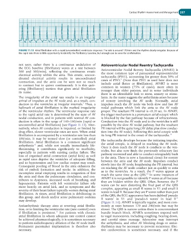
FIGURE 11.13 Atrial fibrillation with a rapid (uncontrolled) ventricular response. The rate is around 170/min and the rhythm clearly irregular. Because of
the rapid rate there is little opportunity to identify the fibrillatory baseline, but enough can be seen for confirmation.
not seen; rather there is a continuous undulation of Atrioventricular Nodal Reentry Tachycardia
the ECG baseline (fibrillatory waves at a rate between Atrioventricular Nodal Reentry Tachycardia (AVNRT) is
300 and 500/min), reflecting the continuous erratic the most common type of paroxysmal supraventricular
electrical activity within the atria. This erratic, uncoor- tachycardia (PSVT), accounting for greater than 50% of
dinated electrical activity results in uncoordinated cases of PSVT. (Note that PSVT as used here does not
5
contraction, and the atria can be seen not so much include atrial flutter or fibrillation). AVNRT is more
to contract but to quiver continuously. It is this quiv- common in women (75% of cases), more often in
ering (fibrillatory) motion that gives atrial fibrillation younger than older patients, and in some individuals
its name.
there is an identifiable link to stress, anxiety or stimu-
The irregularity of the atrial rate results in an irregular lants. As the name suggests the arrhythmia arises because
arrival of impulses at the AV node and, as a result, con- of reentry involving the AV node. Normally, atrial
duction to the ventricles at irregular intervals. Thus, a impulses reach the AV node via both slow and fast AV
7
hallmark of atrial fibrillation is the marked irregularity nodal pathways which link the atria to the AV node
of the ventricular rhythm. The ventricular response rate proper. The resultant PR interval is <0.20 sec. In AVNRT,
to the rapid atrial rate is determined by the state of AV the trigger mechanism is a premature atrial ectopic which
nodal conduction, and in patients with normal AV con- is blocked by the fast pathway because of refractoriness.
duction is often in the range of 140–180/min (rapid or Conduction into the AV node and to the ventricles is still
uncontrolled atrial fibrillation) (see Figure 11.13). Alter- possible by the slow AV nodal pathway, but the resultant
natively, when AV conduction is impaired, or limited by PR interval will be quite long (AV delay plus slow conduc-
drug effect, slower ventricular rates are seen. When atrial tion into the AV node). Following this atrial ectopic with
fibrillation is accompanied by a ventricular rate less than its long PR interval is the onset of the tachycardia. 13
100/min, it may be termed slow (or controlled) atrial
fibrillation. Atrial fibrillation is a common significant The tachycardia develops because the initiating impulse,
arrhythmia and, while not usually immediately life- the atrial ectopic, is delayed in reaching the AV node.
12
threatening, it contributes significantly to morbidity, Once it does reach the AV node it conducts to the ven-
especially in patients with existing cardiac failure. The tricles, but also now finds the previously refractory fast
loss of organised atrial contraction (atrial kick) as well pathway recovered and able to conduct retrogradely back
as rapid rates deprive the ventricles of adequate filling, to the atria. There is now a functional circuit for reentry
and so hypotension and low cardiac output may result. between the atria and the AV node. Impulses conduct
Consequent pooling of blood in the atria enhances the slowly into the AV node, lengthening the PR interval, but
risk of emboli formation and stroke. In addition, the on reaching the AV node conduct just as quickly to atria
incomplete atrial emptying results in congestion of first as to the ventricles. As a result, the P waves appear at
13
the atria and then the pulmonary circulation, and con- much the same time as the QRS. In some instances of
tributes to dyspnoea, increased work of breathing, and AVNRT it is not possible to identify P waves at all because
hypoxaemia. Patients with left ventricular failure rely they are hidden within the QRS. Often, however, the P
more heavily on atrial kick, and so symptoms and the waves can be seen distorting the final part of the QRS
severity of their heart failure typically worsen during atrial complex, appearing as small R waves in V1 and small S
fibrillation. At times, atrial fibrillation is debilitating in waves in lead II. Because they are P waves rather than part
this group, and shock and/or acute pulmonary oedema of the QRS, the ECG appearance has been dubbed ‘pseudo
13
may develop. R waves’ in V1 and ‘pseudo-S waves’ in lead II
(Figure 11.14). AVNRT is typically regular, and most com-
Antiarrhythmic therapy aims at reverting atrial fibrilla- monly at rates between 170 and 240/min but may be
tion, or to limiting the ventricular rate (rate control) even slower. The QRS is narrow unless there is concommitant
12
if fibrillation is persistent. For patients with chronic bundle branch block. AVNRTs sometimes respond well
atrial fibrillation in whom adequate rate control cannot to vagal manoeuvres, including coughing, bearing down,
be achieved pharmacologically, it is sometimes necessary and carotid sinus massage. Adenosine may interrupt
to perform radiofrequency ablation of the AV node itself. the arrhythmia, and other AV blocking drugs or antiar-
Permanent pacemaker implantation is therefore also rhythmics may be necessary to prevent recurrence. Elec-
necessary. tive cardioversion is sometimes necessary, and if the