
Page 281 - ACCCN's Critical Care Nursing
P. 281
258 P R I N C I P L E S A N D P R A C T I C E O F C R I T I C A L C A R E
N N N N S S S S
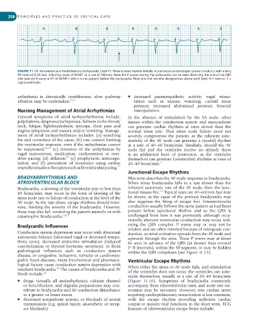
FIGURE 11.14 Atrioventricular Nodal Reentry Tachycardia. Lead V1. There is sinus rhythm initially. A premature atrial ectopic (arrow) conducts with a long
PR interval (0.36 sec), initiating onset of AVNRT at a rate of 140/min. Note the P waves during the tachycardia can be seen distorting the end of the QRS
(the ‘pseudo R wave in V1’ of AVNRT) which is not present before the tachycardia. Note also the monitor designations above each beat: N = normal, S =
supraventricular.
arrhythmia is chronically troublesome, slow pathway ● increased parasympathetic activity: vagal stimu-
ablation may be undertaken. 5,13 lation such as nausea, vomiting, carotid sinus
pressure, increased abdominal pressure, femoral
Nursing Management of Atrial Arrhythmias manipulation.
General symptoms of atrial tachyarrhythmias include: In the absence of stimulation by the SA node, other
palpitations, dyspnoea/tachypnoea, fullness in the throat/ tissues within the conduction system and myocardium
neck, fatigue, lightheadedness, syncope, chest pain and can generate cardiac rhythms at rates slower than the
angina symptoms and nausea and/or vomiting. Manage- normal sinus rate. Thus sinus node failure need not
ment of atrial tachyarrhythmias includes: (a) searching severely compromise the patient, as the inherent auto-
for and correction of the cause; (b) rate control limiting maticity of the AV node can generate a (nodal) rhythm
the ventricular response, even if the arrhythmias cannot at a rate of 40–60 beats/min. Similarly, should the AV
be suppressed; 14,15 (c) reversion of the arrhythmias by node fail and the ventricles receive no stimuli, there
vagal manoeuvres, medication, cardioversion or over- is an additional layer of protection, as the ventricles
16
drive pacing; (d) ablation; (e) prophylactic anticoagu- themselves can generate (ventricular) rhythms at rates of
lation; and (f) prevention of recurrence using cardiac 20–40 beats/min. 7
resynchronisation therapies such as biventricular pacing. 17
Junctional Escape Rhythms
BRADYARRHYTHMIAS AND This term describes the AV node response to bradycardia.
ATRIOVENTRICULAR BLOCK When sinus bradycardia falls to a rate slower than the
Bradycardia, a slowing of the ventricular rate to less than inherent automatic rate of the AV node, then the junc-
7,9
60 beats/min, may occur in the form of slowing of the tional tissues fire. Typical rates are 40–60/min but may
sinus node rate or failure of conduction at the level of the be slower, as the cause of the primary bradycardia may
AV node. As the rate slows, escape rhythms should inter- also suppress the firing of escape foci. In traventricular
vene, limiting the severity of the bradycardia. However, conduction usually follows the same pattern as had been
these may also fail, rendering the patient asystolic or with present before junctional rhythm and so the QRS is
catastrophic bradycardia. 18,19 unchanged from how it was previously, although occa-
sionally aberrant ventricular con duction may occur, wid-
Bradycardic Influences ening the QRS complex. P waves may or may not be
Conduction system depression may occur with abnormal evident and are often inverted because of retrograde con-
duction, as atrial activation spreads from the AV node and
autonomic balance (increased vagal or decreased sympa- upwards through the atria. These P waves may at times
thetic tone), decreased endocrine stimulation (reduced be seen in advance of the QRS (at shorter than normal
catecholamine or thyroid hormone secretion), or from P–R intervals), within the ST segment, or may be hidden
pathological influences such as conduction system within the QRS complexes (see Figure 11.15).
disease, or congestive, ischaemic, valvular or cardiomyo-
pathic heart diseases. Many biochemical and pharmaco- Ventricular Escape Rhythms
logical factors cause conduction system depression with
18
resultant bradycardia. The causes of bradycardia and AV When either the sinus or AV node fails, and stimulation
block include: 18 of the ventricles does not occur, the ventricles can auto-
excite themselves, usually at a rate of 20–40 beats/min
● drugs: virtually all antiarrhythmics, calcium channel (Figure 11.16). Symptoms of bradycardia commonly
or beta-blockers, and digitalis preparations may con- accompany these idioventricular rates, and acute rate res-
tribute to bradycardia and AV conduction disturbance toration may be necessary. However, true cardiac arrest
to a greater or lesser extent requiring cardiopulmonary resuscitation is less common,
● decreased sympathetic activity, or blockade of neural with the escape rhythm providing sufficient cardiac
transmission (e.g. spinal injury, anaesthetic or recep- output to sustain vital functions in the short term. ECG
tor blockade) features of idioventricular escape beats include: