
Page 282 - ACCCN's Critical Care Nursing
P. 282
Cardiac Rhythm Assessment and Management 259
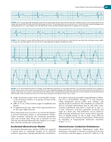
FIGURE 11.15 Sinus bradycardia followed by onset of junctional escape rhythm. Note that the sinus rate is initially around 37/min. It then slows into the
escape rate range of the AV node, which then discharges at 35/min. The junctional beats are not preceded by P waves: the more slowly discharging sinus
node probably has its P waves hidden first in the QRS of the second-last beat and then distorts the ST segment of the last beat.
FIGURE 11.16 Ventricular escape rhythm (idioventricular rhythm). Note that after the first sinus beat, the slow rate allows the ventricular escape rhythm
to emerge. The resultant rhythm is at a rate of 35/min, with wide QRS complexes and absent P waves.
I I
I I
FIGURE 11.17 Accelerated Idioventricular Rhythm (AIVR) following reperfusion in myocardial infarction. An accelerated ventricular focus emerges at
65/min, taking over from the slower sinus rate of 60/min. It then accelerates gradually until settling at a rate of 85/min by the end of the second strip. This
display of rate ‘warm-up’ at onset is a characteristic of arrhythmias due to increased automaticity. The distortion of the ST segment from the third beat of
AIVR onwards is due to retrograde conduction to the atria, and explains the absence of the sinus P waves.
● single ventricular ectopic beats occurring after a pause thus often indicating successful revascularisation follow-
in the dominant rhythm, or as groups of beats at the ing PCI or thrombolytic therapy. 20,21 It may therefore
slow escape rate imply therapeutic success rather than mishap, and
● QRS >0.12 sec, often notched, larger in amplitude and usually needs no treatment. The arrhythmia is commonly
bizarre due to increased automaticity and as with other auto-
● ST segment and T wave, often in the opposite direction maticity arrhythmias may show a ‘warm-up’ in rate, i.e.
to the major QRS direction. it may commence and then gradually accelerate and
settle at a faster rate. This behaviour can be useful in
When these beats occur at a rate of 20–40/min the rhythm differentiating arrhythmias from reentry which typically
is termed ventricular escape, or idioventricular rhythm. have an abrupt change in rate as their onset. When it
Under excitatory influences the ventricular pacemaker occurs outside of the context of reperfusion, AIVR should
cells may increase their firing rate to between 60 and be regarded as inappropriate ventricular excitation
100/min (accelerated idioventricular rhythm) or to faster (Figure 11.17).
than 100/min (ventricular tachycardia). 20
Accelerated Idioventricular Rhythm Atrioventricular Conduction Disturbances
Accelerated idioventricular rhythm (AIVR) has assumed Atrioventricular conduction disturbances make their
a special place in cardiology because of its relatively appearance as delayed or blocked conduction from atria
common appearance during postinfarction reperfusion, to ventricles, and thus appear as altered P–QRS (or P–R)