
Page 324 - ACCCN's Critical Care Nursing
P. 324
Cardiac Surgery and Transplantation 301
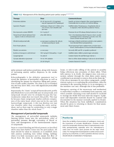
TABLE 12.2 Management of the bleeding patient post-cardiac surgery 21,22,25,33,34,44
Therapy Dose Comments/issues
Protamine sulphate 25–50 mg slow IV (<10 mg/min); Specific antidote to heparin. May cause hypotension.
may be repeated if ACT prolonged Contraindicated in patient with seafood allergy.
Aprotinin (Trasylol) continuous infusion of 2 million units Antifibrinolytic. Proteinaceous. Anaphylaxis risk on
over 30 min, then 500,000 units re-exposure. Alert should be posted on history.
per hour
Desmopressin acetate (DDAVP) 0.4 mcg/kg IV Promotes factor VIII release; limited evidence for use.
‘Pump blood’ (blood retrieved from often 400–800 mL This is the remaining blood in bypass circuit; usually
bypass circuit at end-operation) centrifuged before returning to patient; note: this
blood contains heparin from CPB.
Whole blood/packed cells as necessary to achieve Hb >80 g/L Autologous blood sometimes available when patients
or more according to needs have donated blood preoperatively.
Fresh frozen plasma as necessary ‘Broad-spectrum’ factor replacement; contains most
factors. Useful adjunct to massive blood transfusion.
Platelet concentrates as necessary Generally ABO and Rh compatible preferred.
Epsilon-aminocaproic acid (Amicar) 100 mg/kg IV followed by 1–2 g/h Antifibrinolytic. Inhibits plasminogen activation.
Cryoprecipitate 10 units IV Contains factor VIII and fibrinogen (factor I).
Calcium chloride or gluconate 10 mL 10% solution Used to offset citrate binding of calcium in stored blood.
Prothrombinex 20–50 IU/kg IV Contains factors II, IX and X.
pulse pressure and pulsus paradoxus, along with features loops, or side-to-side rolling of the patient to possibly
of increasing anxiety and/or dyspnoea in the awake bring collections into proximity of drain tubes. When
patient. tube patency is in doubt, the surgeon may even pass a
suction catheter through the chest drain under aseptic
Echocardiography is the definitive assessment tool to conditions in an attempt to remove clots at the drain tip.
23
reveal the presence of pericardial collections as well as If the above measures do not relieve tamponade, consid-
identifying the impact on relaxation, filling and contrac- eration is given to re-exploring the pericardium, either by
tion of each cardiac chamber. The chest X-ray is of limited returning to the operating theatre or, in an emergency, to
use and may show little, even with significant pericardial the intensive care unit, although this is less preferable.
collections.
Emergency opening of the sternotomy and mediastinal
Importantly, the ‘classic’ or typical haemodynamic profile re-exploration requires a coordinated team response, and
described above is not uniformly seen in tamponade, and where possible operating room staff should be included
tamponade should never be excluded because the hae- to manage the sterile field and assist the surgeon. Equip-
modynamic status does not match this profile. This may ment and disposable materials should be counted and
be because classic tamponade implies uniform compres- documented in the manner normally applied in theatre.
sion of the entire heart, which may not be the case with When the situation has been stabilised, consideration
haemorrhagic tamponade. A clot may develop over just should be given to returning to theatre for final assess-
one chamber rather than occupying the entire pericar- ment and chest closure.
dium, and so there may be compromise to only a single
chamber rather than the whole heart. 21,23
Management of pericardial tamponade
The management of pericardial tamponade includes
limiting further losses into the pericardium, relief of Practice tip
pericardial pressure through evacuation of blood or
clots, and management of the haemodynamic impact Given the variability of presentation of cardiogenic shock, and
of tamponade. the importance of accurate identification, clinicians should
search for tamponade whenever there is haemodynamic insta-
Steps to control bleeding and blood pressure as described bility postoperatively, especially when the haemodynamic
above may limit further losses into the pericardium. All status does not match classic patterns for the major shock
steps should be taken to maintain or re-establish chest states. The management of postoperative cardiac arrest accom-
tube patency (crushing clots within tubing, ‘milking’ panying any arrhythmia, as well as pulseless electrical activity,
when it is truly necessary) and to ensure free flow of should include consideration of tamponade.
blood from the chest by avoiding dependent tubing