
Page 326 - ACCCN's Critical Care Nursing
P. 326
Cardiac Surgery and Transplantation 303
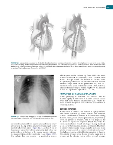
FIGURE 12.4 Intra-aortic balloon catheter. On the left the inflated catheter can be seen behind the heart, with its tip below the arch of the aorta and the
left subclavian artery. The balloon cycles between inflated (during diastole), and deflated (during systole) as on the right above. Blood fills the aorta while
the balloon is deflated, and with inflation the balloon almost fills the descending aorta, displacing 40 mL blood from the aorta to the coronary and systemic
circulation. (Courtesy Datascope Corporation, Fairfield, NJ).
which opens at the catheter tip from which the aortic
pressure waveform is monitored; and a helium drive
lumen, through which the helium is shuttled from
the pumping console to the catheter balloon. Balloon
volumes range from 25 mL (paediatric use) and 34–
50 mL in adults (most commonly used is 40 mL balloon)
and selected according to patient height (40 mL balloon
is used for a patient height of 162–183 cm).
PRINCIPLES OF COUNTERPULSATION
When pumping is initiated, the balloon will be
inflated rapidly at the onset of diastole of each cardiac
cycle and then deflated immediately just before the
onset of the next systole; this sequence is referred to as
counterpulsation.
Balloon Inflation
At the onset of diastole, the balloon is rapidly inflated
with (most commonly) 40 mL helium. This inflation
causes a sudden rise in pressure in the aortic root during
FIGURE 12.5 IABP catheter position in CXR, the tip is located in second
intercostal space anterior ribs or fifth intercostal space posterior ribs. diastole, raising mean arterial pressure and, importantly,
coronary perfusion pressure. The blood displaced by
the balloon expansion improves blood flow into the
commonly advanced from a percutaneous femoral artery coronary circulation (which fills largely during diastole),
access until the tip of the catheter is situated just below as well as to the brain and systemic circulation. Thus there
the left subclavian artery (Figure 12.4). A chest X-ray or is improved myocardial oxygen supply, increased mean
46
fluoroscopy should reveal the catheter tip just below the arterial pressure, as well as improved systemic perfusion.
aortic arch, or at the level of the second anterior intercos- The balloon remains inflated for the duration of diastole.
tal space or fifth posterior intercostal space (Figure 12.5). The arterial pressure wave should reveal a sharp rise in
The catheter has two lumens – a monitoring lumen, pressure at the dicrotic notch, with a second pressure