
Page 357 - ACCCN's Critical Care Nursing
P. 357
334 P R I N C I P L E S A N D P R A C T I C E O F C R I T I C A L C A R E
From interruption to these vital cellular activities is a reduction
pulmonary Airway in organ or tissue function, which in turn compromises
artery Impaired system and body functions.
ventilation
Alveolus Changes to the oxyhaemoglobin dissociation curve also
occur in states related to hypoxia. The curve shifts to the
right when there is acidosis and/or raised levels of PCO 2
as commonly seen in respiratory failure. Although this
Alveolocapillary change may alter patient oxygen saturation readings, the
membrane To Hypoxaemia increased release of oxygen from haemoglobin to the
Normal V/Q pulmonary vein Low V/Q tissues has obvious benefits for tissue oxygenation and
cellular metabolism. 7
Blocked Impaired perfusion Compensatory Mechanisms to
ventiation Optimise Oxygenation
Alveolar
Collapsed dead space When PO 2 in the alveolus is reduced, hypoxic pulmonary
alveous
vasoconstriction occurs, with contraction of smooth
muscles in the small arterioles in the hypoxic region,
directing blood flow away from the hypoxic area of the
lung. Peripheral chemoreceptors also detect hypoxaemia
7
and initiate compensatory mechanisms to optimise cel-
Hypoxaemia Hypoxaemia lular oxygen delivery. Initial responses are increased respi-
Shunt (very low) V/Q High V/Q ratory rate and depth of breathing, resulting in increased
minute ventilation, and raised heart rate with possible
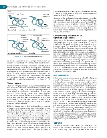
FIGURE 13.11 Ventilation perfusion mismatch.
18
vasoconstriction as the body attempts to maintain oxygen
delivery and uptake. This overall up-regulation cannot be
an overall reduction in blood oxygen levels, which can sustained indefinitely, particularly in a person who is
usually be countered by compensatory mechanisms. 1
critically ill, and compensatory mechanisms begin to fail
Intrapulmonary shunting is an extreme case of V/Q mis- with worsening hypoxaemia and cellular and organ dys-
match. Shunting occurs when blood passes alveoli that function. Unless the hypoxaemia is reversed and/or respi-
are not ventilated. There can be significant intrapulmo- ratory and cardiovascular support is provided, irreversible
nary shunting, and therefore overwhelming reductions in hypoxia and death will ensue.
18
PaO 2 . Carbon dioxide levels may still be normal but
depending on the onset and progression of the respira- INFLAMMATION
tory pathophysiology, compensatory mechanisms may Inflammatory processes can occur at a local level (e.g. as
not be able to maintain homeostasis 1,11 (see Figure 13.11). a result of inhalation injuries, aspiration or respiratory
infections) or are secondary to systemic events (e.g. sepsis,
Tissue Hypoxia trauma). Damage to the pulmonary endothelium and
There are few physiological changes with mild hypoxae- type I alveolar cells appear to play a key role in the inflam-
20
mia (when O 2 saturation remains at 90% despite a PaO 2 matory processes associated with ALI. Once triggered,
of 60 mmHg [8 kPa]), with only a slight impairment in inflammation results in platelet aggregation and comple-
mental state. If hypoxaemia deteriorates and the PaO 2 ment release. Platelet aggregation attracts neutrophils,
drops to 40–50 mmHg (5.3–6.7 kPa), severe hypoxia which release inflammatory mediators (e.g. proteolytic
of the tissues ensues. Hypoxia at the central nervous enzymes, oxygen free radicals, leukotrienes, prostaglan-
system level manifests with headaches and somnolence. dins, platelet-activating factor [PAF]). Neutrophils also
Compensatory mechanisms include catecholamine appear to play a key role in the perpetuation of ALI/
1
release, and a decrease in renal function results in sodium ARDS. As well as altering pulmonary capillary permea-
retention and proteinuria. 19 bility, resulting in haemorrhage and fluid leak into the
pulmonary interstitium and alveoli, mediators released
Different tissues vary in their vulnerability to hypoxia,
with the central nervous system and myocardium at most by neutrophils and some macrophages precipitate pul-
risk. Hypoxia in the cerebral cortex results in a loss of monary vasoconstriction. Resulting pulmonary hyperten-
function within 4–6 seconds, loss of conscious in 10–20 sion leads to diminished perfusion to some lung areas,
seconds and irreversible damage in 3–5 minutes. In an with dramatic alterations to both perfusion and ventila-
11
environment that lacks oxygen, cells function by anaero- tion leading to significant V/Q mismatches, and the sub-
bic metabolism and produce much less energy (adenos- sequent signs and symptoms typically seen in patients
ine triphosphate [ATP]) than with aerobic metabolism with pulmonary inflammation/oedema.
(2 versus 38 ATP molecules per glucose molecule), and
lactic acid increases. With less available energy, the effi- OEDEMA
+
+
ciency of cellular functions such as the Na /K pump, Pulmonary oedema also alters gas exchange, and
nerve conduction, enzyme activity and transmembrane results from abnormal accumulation of extravascular
receptor function diminishes. The overall effect of fluid in the lung. The two main reasons for this are:
19