
Page 363 - ACCCN's Critical Care Nursing
P. 363
340 P R I N C I P L E S A N D P R A C T I C E O F C R I T I C A L C A R E
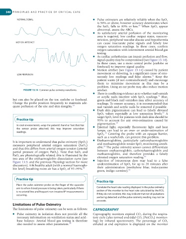
NORMAL SIGNAL ● Pulse oximeters are relatively reliable when the SaO 2
is 90% or above, however accuracy deteriorates when
33
the SaO 2 falls to 80% or less. When SpO 2 appear
abnormal, assess the ABGs.
● As satisfactory arterial perfusion of the monitoring
area is required, low cardiac output states, vasocon-
striction, peripheral vascular disease and hypothermia
MOTION ARTEFACT can cause inaccurate pulse signals and falsely low
oxygen saturation readings. In these cases, confirm
oxygen saturation with intermittent arterial blood gas
testing.
● As cardiac arrhythmias can impair perfusion and flow,
signal quality may be compromised (see Figure 13.14).
In these cases, use a more central probe (earlobe or
forehead) to improve signal quality.
● Motion artefact (see Figure 13.14) caused by patient
LOW PERFUSION movement or shivering, is a significant cause of erro-
36
neously low readings and false alarms. Keep the
patient warm (if not contraindicated) and encourage
them to minimise movement as this may be a
problem. Using an ear probe may also reduce motion
FIGURE 13.14 Common pulse oximetry waveforms. artefact.
● There is conflicting evidence as to whether nail varnish
36
or acrylic nails interfere with SpO 2 readings. Blue,
but can also be placed on the toe, earlobe or forehead. green and black nail varnishes may affect accuracy of
Change the probe position frequently to maintain ade- readings. To ensure accuracy, it is recommended that
quate perfusion of the site and skin integrity. 32 nail varnish and acrylic nails be removed if possible.
● Dark skin pigmentation can lead to falsely elevated
37
SpO 2 values especially at low saturation levels. A
target SpO 2 level for patients with dark skin should be
Practice tip 95% to account for any over-estimation caused by
pigmentation. 33
In cool environments, wrap the patient’s hand or foot that has
the sensor probe attached; this may improve saturation ● External light, especially fluorescent light and heat
readings. lamps, can lead to an over- or under-estimation of
35
SpO 2 . Covering the probe with an opaque barrier,
such as a washcloth, can prevent this problem.
● Dyshaemoglobins, particularly carboxyhaemoglobin
It is important to understand that pulse oximetry (SpO 2 )
measures peripheral arterial oxygen saturation (SaO 2 ) and methaemoglobin render SpO 2 monitoring unreli-
33
and that this differs from arterial oxygen tension (arterial able. The pulse oximetry sensor cannot differentiate
partial pressure of oxygen; PaO 2 ). Note that SaO 2 and between oxyhaemoglobin, carboxyhaemoglobin and
PaO 2 are physiologically related; this is illustrated by the methaemoglobin, and therefore provides a falsely
35
two axes of the oxyhaemoglobin dissociation curve (see elevated oxygen saturation reading.
Figure 13.9, and the previous Physiology section for more ● Injection of intravenous dyes may lead to a false
discussion). A fit healthy adult (with a normal haemoglo- underestimation of SpO 2 for up to 20 minutes after
bin level) breathing room air has a SpO 2 of 97–99%. 34 their administration (methylene blue, indocyanine
green, indigo carmine). 33
Practice tip
Practice tip
Place the pulse oximeter probe on the finger of the opposite
arm to where blood pressure is being taken, particularly if there Correlate the heart rate reading displayed in the pulse oximetry
is no arterial line and frequent non-invasive BP measurement is section of the monitor to the heart rate calculated by the ECG.
occurring. If they do not correlate, this may indicate that not all pulsations
are being detected and the pulse oximetry reading may not be
accurate.
Limitations of Pulse Oximetry
The limitations of pulse oximetry can be seen as follows: CAPNOGRAPHY
● Pulse oximetry in isolation does not provide all the Capnography monitors expired CO 2 during the respira-
necessary information on ventilation status and acid– tory cycle (also termed end-tidal CO 2 [PetCO 2 ] monitor-
base balance. Arterial blood gas testing is therefore ing) by infrared spectrometry. The percentage of CO 2
also needed to assess other parameters. 35 exhaled at end expiration is displayed on the monitor