
Page 310 - Concise Pathology for Exam Preparation ( PDFDrive )
P. 310
12 Haematology 295
Characteristic Features
• Affects middle-aged to elderly individuals who present with insidious onset moderate
to severe anaemia.
• The peripheral smear shows a dimorphic picture with microcytic, hypochromic and
normocytic cells.
• Iron overload with characteristic ringed sideroblasts are seen in the bone marrow (iron
enters mitochondria surrounding the nucleus, cannot exit and appears as ‘rings’ with
Prussian blue staining).
• Siderocytes called Pappenheimer bodies (containing nonhaem iron granules) are seen
in the RBCs.
• Bone marrow shows erythroid hyperplasia and ineffective erythropoiesis. Dyserythro-
poiesis is common.
• There is marked increase in serum iron and transferrin saturation.
Q. Enumerate the causes of microcytic hypochromic anaemia.
Ans. Causes of microcytic hypochromic anaemia include
• Iron-deficiency anaemia
• Thalassaemia syndromes
• Sideroblastic anaemia
• Chronic lead poisoning
• Some cases of anaemia associated with chronic disorders
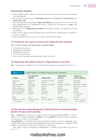
Q. Enumerate the salient features of hypochromic anaemias.
Ans. Salient features of different types of hypochromic anaemias are summarized in Table 12.5.
TABLE 12.5. Salient features of different hypochromic anaemias
Iron deficiency Sideroblastic Anaemia of
Features anaemia Thalassaemia anaemia chronic disorders
Serum iron Decreased Normal or increased Increased Decreased
TIBC Raised Normal Normal Decreased
Percent saturation Decreased Normal to increased Normal to increased Decreased to normal
Serum ferritin Decreased Normal Normal Normal to increased
Marrow iron stores Absent Present Present Present
Iron in normoblasts Absent Present Ring sideroblasts Absent
Hb electrophoresis Normal Abnormal Normal Normal
Red cell indices Reduced Very low Low Low normal to
reduced
RDW High Normal Normal Normal
Diagnostic feature/ Decreased serum Hb electrophoresis Presence of ring Presence of normo-
investigation ferritin sideroblasts cytic population
Q. Describe the aetiopathogenesis, clinical features and haematologic
picture of macrocytic anaemia.
Ans. A macrocyte is defined as a large red cell with increased volume (MCV . 100 fL),
diameter (. 8.5 mL) and thickness (MCH .31.5 pg; MCHC normal). Increased thickness
is perceived as a loss of central pallor.
A megaloblast is so labelled when a large erythroid cell shows nuclear-cytoplasmic asyn-
chrony (maturation of nucleus lags behind maturation of cytoplasm due to impaired DNA
synthesis caused by the deficiency of vitamin B 12 and folate).
The salient features pertaining to metabolism of vitamin B 12 and folate are listed in
Table 12.6.
mebooksfree.com