
Page 308 - Concise Pathology for Exam Preparation ( PDFDrive )
P. 308
12 Haematology 293
• Tongue: Atrophy of papillae, shiny or glazed tongue, glossitis and angular stomatitis
• Plummer–Vinson (Paterson–Kelly) syndrome: Characterized by chronic iron defi-
ciency, dysphagia and glossitis; seen in middle-aged to elderly women who have
chronic iron deficiency and a fine web or band composed of desquamating epithelial
cells at the oesophageal entrance (postcricoid web). These patients present with dys-
phagia to solids.
• Pica: This is defined as a craving to eat substances like dirt, clay, salt, hair and is a
typical manifestation of iron deficiency.
• Recurrent infections: Iron deficiency induces defective lymphocyte-mediated immu-
nity and impairs bacterial killing by phagocytes leading to impaired immunity and re-
current infections.
Laboratory Diagnosis
1. General blood parameters
(a) Hb: Decreased
(b) RBC count: Decreased
(c) RBC indices: Reduced/low
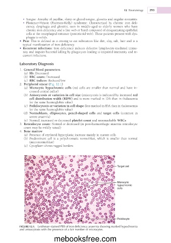
2. Peripheral smear (Fig. 12.1)
(a) Microcytic hypochromic cells (red cells are smaller than normal and have in-
creased central pallor)
(b) Anisocytosis or variation in cell size (anisocytosis is indicated by increased red
cell distribution width (RDW) and is more marked in IDA than in thalassemia
for the same haemoglobin value)
(c) Poikilocytosis or variation in cell shape (less marked in IDA than in thalassaemia
for the same haemoglobin value)
(d) Normoblasts, elliptocytes, pencil-shaped cells and target cells (common in
severe anaemia)
(e) Normal, increased or decreased platelet count and unremarkable WBCs
3. Reticulocyte count: Normal or decreased (in post-haemorrhagic anaemia reticulocyte
count may be mildly raised)
4. Bone marrow
(a) Presence of erythroid hyperplasia; increase mainly in mature cells
(b) Predominant cell is a polychromatic normoblast, which is smaller than normal
(micronormoblast)
(c) Cytoplasm shows ragged borders
Target cell
Microcytic
hypochromic
cells
Poikilocyte
FIGURE 12.1. Leishman-stained PBS of iron deficiency anaemia showing marked hypochromia
and anisocytosis with the presence of a fair number of microcytes.
mebooksfree.com