
Page 305 - Concise Pathology for Exam Preparation ( PDFDrive )
P. 305
290 SECTION II Diseases of Organ Systems
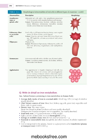
TABLE 12.4. Morphological abnormalities of red cells in different types of anaemias—cont’d
Abnormalities Description Morphology
Acanthocytes Spheroidal red cells with a few spiny/thorny projections
(spur or (5–10 in number with irregular thickness and spacing)
spicule cells) Causes: Post-splenectomy, alcoholic cirrhosis, pyruvate
kinase deficiency, autoimmune haemolytic anaemia
(AIHA), severe burns and renal disease
Echinocytes (Burr Small cells or cell fragments bearing shorter, more regular
or sea urchin spines 10–30 in number, evenly spaced
cells) Causes: Uraemia, pyruvate kinase deficiency, blood stor-
age, ATP depletion, calcium accumulation and contact
with glass
Elliptocytes Elliptical cells
Causes: Hereditary elliptocytosis, thalassaemia, sickle cell
trait, iron deficiency, megaloblastic and myelophthisic
anaemia
Stomatocytes Uniconcave red cells with a slit-like area of central pallor
Causes: Hereditary stomatocytosis, alcoholism, cirrhosis,
obstructive liver disease
Agglutination True agglutination is irregular clumping of red cells into
grape like clusters (in contrast with pseudoagglutina-
tion or rouleaux formation which is observed in para-
proteinaemias, hypergammaglobulinaemia and fibrino-
genaemia which causes stacking of red cells like coins)
Q. Write in detail on iron metabolism.
Ans. Salient features pertaining to iron metabolism in human body:
• Average daily intake of iron in a normal adult: 10–20 mg; 10% (1-2 mg) of which is
usually absorbed.
• Chief dietary sources of iron: Meat, liver, kidney, egg yolk, green leafy vegetables and
fruits; milk is a poor source.
• Dietary iron: Two major types:
Haem iron (found in animal products and more readily absorbed)
Nonhaem iron (found as inorganic iron in vegetables, less readily absorbed)
• Total body iron: 3–5 g (proportionate to body weight)
• Eighty percent of functional iron is in haemoglobin: 2–3 g
• Storage or available tissue iron (ferritin and hemosiderin): 1 g
• Essential or nonavailable iron (myoglobin and other enzymes of cellular respiration):
0.5 g
• Iron is transported in the plasma, bound to a glycoprotein called transferrin (plasma
or transport iron): 0.003–0.004 g
mebooksfree.com