
Page 320 - Concise Pathology for Exam Preparation ( PDFDrive )
P. 320
12 Haematology 305
Laboratory Evidence of Damage to RBCs
• Presence of fragments of red cells (schistocytes) and spherocytes in the peripheral smear
• Positive direct Coombs test, if haemolysis is immunological in origin
• Shortened red cell life (decreased to 30–40 days; normal 120 days)
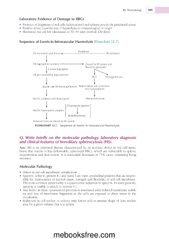
Sequence of Events in Intravascular Haemolysis (Flowchart 12.7)
Breakdown
Hb monomers and dimers Hb tetramers
Hb-haptoglobin complex Cleared by RE system and
filtered by glomeruli
↓ Serum haptoglobin
Hb gets oxidized in large amounts
PCT Hemoglobinuria
MetHb Methaemoglobinuria Reabsorption and conversion
into haemosiderin
MetHb combines with haemopexin Haemosiderinuria
If haemopexin depleted
MetHb–haemopexin complex
Methalbuminemia
Removed from circulation by RE system
FLOWCHART 12.7. Sequence of events in intravascular haemolysis.
Q. Write briefly on the molecular pathology, laboratory diagnosis
and clinical features of hereditary spherocytosis (HS).
Ans. HS is an inherited disease characterized by an intrinsic defect in red cell mem-
brane that results in less deformable, spheroidal RBCs, which are vulnerable to splenic
sequestration and destruction. It is autosomal dominant in 75% cases; remaining being
recessive.
Molecular Pathology
• Defect in red cell membrane cytoskeleton
• Spectrin, ankyrin, protein 4.1 and band 3 are main cytoskeletal proteins that are respon-
sible for maintenance of normal shape, strength and flexibility of red cell membrane.
The most common abnormality is a quantitative reduction in spectrin. In some patients,
spectrin is unable to attach to protein 4.1.
• Any defect in these cytoskeleton proteins is associated with reduced membrane stabil-
ity and loss of membrane fragments as the cells are exposed to shear stress in the
circulation.
• Reduction in cell surface to volume ratio forces cells to assume shape of least surface
area for a given volume that is a sphere.
mebooksfree.com