


Page 36 - Concise Pathology for Exam Preparation ( PDFDrive )
P. 36
1  Cell Injury and Cell Death  21


                   the phosphatidylserine so that it is expressed on the outer membrane and is easily
                   recognized by the macrophage.
                 (b)  Secretion of soluble factors by apoptotic cells, eg, thrombospondin, which recruit
                   macrophages.
                 (c)  Coating of apoptotic cells by natural antibodies and proteins of the complement
                   system, which are easily recognized by macrophage receptors.

             Q. Differentiate between apoptosis and necrosis.
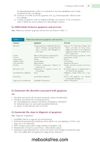
             Ans. Differences between apoptosis and necrosis are shown in Table 1.7.


               TABLE 1.7.   Differences between apoptosis and necrosis
               Features              Apoptosis                      Necrosis
               Definition            Programmed and coordinated cell death,   Spectrum  of  morphologic  changes  that
                                      which  eliminates  unwanted/harmful   follow  cell  death  in  living  tissue,
                                      cells  or  removes  cells  damaged  be-  largely resulting from the progressive
                                      yond repair                     degradative action of enzymes on le-
                                                                      thally injured cells
               Causes                May be physiological or pathological  Always pathological, eg, hypoxia, toxins
               Involves              Single or small groups of cells  Large groups of cells
               Inflammation          Absent                         Present
               Cellular change       Cell shrinkage                 Cell swelling
               Cell membrane         Bleb formation                 Membrane disruption
               Nucleus               Chromatin  condensation  followed  by   Nuclear pyknosis, karyolysis and kary-
                                      fragmentation                   orrhexis
               Removal of cell       Phagocytosis  of  apoptotic  bodies  by   Enzymatic digestion or phagocytosis of
                                      macrophages                     cell debris by macrophages
               Lysosomes/other organelles  Intact                   Hydrolytic  enzyme  release  due  to  rup-
                                                                      ture
               Mechanism             Genetically coordinated        Due  to  ATP  depletion,  free  radicals,
                                                                      mitochondrial damage, etc.
               Agarose gel electrophoresis  Stepladder DNA pattern  Diffuse DNA pattern


             Q. Enumerate the disorders associated with apoptosis.
             Ans.

               1.  Disorders associated with decreased apoptosis: cancer, autoimmunity
               2.  Disorders associated with increased apoptosis:
                 (a)  Neurodegenerative diseases (Alzheimer, Huntington, Parkinson)
                 (b)  Ischaemic injury in stroke and myocardial infarction
                 (c)  Death of virus-infected cells as in AIDS


             Q. Enumerate the steps in diagnosis of apoptosis.
             Ans. Diagnosis of apoptosis:

               1.  Stepladder pattern on agarose gel electrophoresis
               2.  Terminal deoxynucleotidyl transferase biotin-dUTP nick end labelling (TUNEL) tech-
                nique for in vivo detection
               3.  H&E, Feulgen and acridine orange staining of apoptotic cells
               4.  Measurement of cytosolic cytochrome c and activated caspase
               5.  Expression of phosphatidylserine on the outer leaflet of the plasma membrane by apop-
                totic cells enables their recognition by using the dye Annexin V


                                  mebooksfree.com

