


Page 40 - Concise Pathology for Exam Preparation ( PDFDrive )
P. 40
1  Cell Injury and Cell Death  25


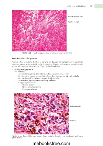
                                                                      Smooth muscle cells

                                                                      Hyaline change


                       FIGURE 1.15.  Hyaline degeneration in a leiomyoma (H&E; 2003).

             Accumulation of Pigments
             Pigment refers to material that has colour and can be seen without staining. In pathology,
             pigments play an important role in the diagnosis of diseases such as gout, jaundice, mela-
             nomas, albinism and haemorrhage. They can be classified as
               1.  Endogenous pigments
                 (a)  Melanin:
                     (i)  Nonhaemoglobin-derived brown-black pigment (Fig. 1.16)
                    (ii)  Normally present in skin, hair, choroids, meninges and adrenal medulla
                     (iii)  Synthesized by melanocytes and dendritic cells
                   Disorders of pigmentation involving melanin:
                   •  Hyperpigmentation:
                     •  Addison disease
                     •  Adrenogenital syndrome
                     •  Chloasma/melasma


                                                                        Malignant cells


                                                                        Melanin


             FIGURE  1.16.  Intracellular  and  extracellular  melanin  deposits  in  a  malignant  melanoma
             (H&E; 4003).


                                  mebooksfree.com

