


Page 37 - Concise Pathology for Exam Preparation ( PDFDrive )
P. 37
22    SECTION I  General Pathology

                     Q. Classify intracellular accumulations and write briefly about them.

                     Ans. Intracellular accumulations include the following:

                     Accumulation of Normal Cellular Constituent in Excess
                       1.  Water
                         (a)  Cloudy swelling
                            (i)  A form of reversible injury, cloudy swelling is also called granular degeneration
                               (named so because of the presence of prominent protein granules in the cyto-
                               plasm)
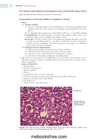
                            (ii)  It commonly affects hepatocytes, renal tubular cells (Fig. 1.13) and myocardium
                           Gross pathology: The affected organ is enlarged, soft and pale (pallor is due to me-
                             chanical compression of capillaries by retained water).
                           Microscopy: Cells are swollen, full of proteinaceous granules (thought to be frag-
                             mented mitochondrial proteins or products of disturbed protein metabolism),
                             and have frayed cell margins. Nuclei are normal in early stages, but could later
                             appear faint or intensely staining.
                         (b)  Hydropic/vacuolar degeneration
                             (i)  This is an extension of changes seen in cloudy swelling
                            (ii)  Affected cells are ballooned, pale, watery and vacuolated
                             (iii)  Vacuoles coalesce and push nucleus towards the periphery
                            (iv)  Cell bursts and nucleus undergoes karyorrhexis/lysis
                       2.  Fat:  Abnormal  accumulation  of  triglycerides  in  the  cytosol  of  parenchymal  cells  is
                        called fatty change (steatosis). It mainly affects liver and heart but can also be seen in
                        muscle and kidney.
                        Causes of fatty liver
                        •  Alcohol abuse.
                        •  Starvation/malnutrition.
                        •  Diabetes mellitus.
                        •  Obesity.
                        •  Hepatotoxins like CCl 4 , ether, aflatoxins.
                        •  Certain drugs, like steroids, tetracycline and aspirin (Reye syndrome).
                        •  Hypoxia in anaemia and cardiac failure.
                        •  Late pregnancy.
                        •  Chronic illness, like tuberculosis.


                                                                                Glomerulus


                                                                                Tubules lined by
                                                                                swollen cells with
                                                                                frayed margins


                     FIGURE 1.13.  Section from cloudy swelling kidney showing tubules lined by swollen cells
                     with frayed margins and increased granularity (H&E; 2003).


                                  mebooksfree.com

