


Page 39 - Concise Pathology for Exam Preparation ( PDFDrive )
P. 39
24    SECTION I  General Pathology


                                                                             Hepatocytes showing
                                                                             large cytoplasmic
                                                                             vacuoles


                                                                             Eccentric nuclei


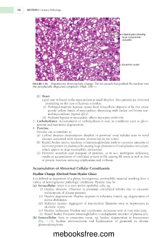
                     FIGURE 1.14.  Hepatocytes showing fatty change. The fat vacuole has pushed the nucleus into
                     the peripherally displaced cytoplasm (H&E; 2003).


                       (b)  Heart
                           Lipid may be found in the myocardium as small droplets. Two patterns are observed
                             depending on the type of hypoxic stimulus:
                            (i)  Prolonged moderate hypoxia: causes focal intracellular deposits of fat that create
                               grossly yellow bands of myocardium alternating with darker red-brown nor-
                               mal myocardium (tigered effect).
                             (ii)   Profound hypoxia or myocarditis: affects myocytes uniformly.
                       3.  Carbohydrates: Accumulation of carbohydrates is seen in conditions such as glyco-
                        genosis and mucinous degeneration.
                       4.  Proteins:
                        Proteins can accumulate as
                        (a)  Colloid  droplets  (reabsorption  droplets  in  proximal  renal  tubules  seen  in  renal
                           diseases associated with excessive protein loss in the urine).
                       (b)  Russell bodies (active synthesis of immunoglobulins leads to excessive amounts of
                           secretory protein in plasma cells causing huge distension of endoplasmic reticulum,
                           which appear as large eosinophilic inclusions).
                        (c)  Defective  secretion  and  transport  of  proteins,  as  in  a-1  antitrypsin  deficiency,
                           results in accumulation of misfolded protein in ER causing ER stress as well as loss
                           of protein function inducing emphysema and cirrhosis.

                     Accumulation of Abnormal Cellular Constituents

                     Hyaline Change (Derived From Hyalos Glass)
                     It is defined as deposition of a glassy, homogenous, eosinophilic material resulting from a
                     variety of heterogeneous pathologic conditions. Hyaline may be
                     (a)  Intracellular: when it is seen within epithelial cells, eg,
                           (i)  Hyaline  droplets:  Observed  in  proximal  convoluted  tubules  due  to  excessive
                             reabsorption of plasma proteins.
                          (ii)  Hyaline degeneration: Hyaline deposits in voluntary muscle, eg, degeneration of
                             rectus abdominis.
                           (iii)  Mallory’s  hyaline:  Aggregates  of  intermediate  filaments  seen  in  hepatocytes  in
                             alcoholic injury.
                          (iv)  Hyaline inclusions: Nuclear and cytoplasmic inclusions seen in viral infections.
                          (v)  Russell bodies: Excessive immunoglobulins in endoplasmic reticulum of plasma cells.
                       (b)  Extracellular:  Seen  in  connective  tissue,  eg,  hyaline  degeneration  in  leiomyomas
                         (Fig.  1.15),  hyaline  arteriosclerosis  and  hyalinization  of  glomeruli  in  chronic
                         glomerulonephritis.


                                  mebooksfree.com

